Characterization
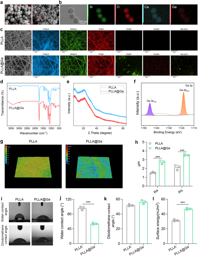
SEM and TEM have been utilized to characterize the morphology and elemental composition of the synthesized Ga-MBG. As depicted in Fig. 1a, Ga-MBG seems as uniform spherical particles with a mean diameter of roughly 100 nanometers, in step with TEM observations. TEM pictures (Fig. 1b) reveal the uniform spherical morphology of Ga-MBG particles with a mean diameter of roughly 100 nm. As well as, TEM picture clearly exhibits the mesoporous construction of Ga-MBG. Vitality-dispersive X-ray spectroscopy (EDS) mapping confirms the homogeneous distribution of Ga inside the MBG framework, indicating profitable doping of Ga ions. As demonstrated in Fig. 1c, the profitable preparation of Ga-MBG loaded PLLA electrospun fibers resulted within the uniform distribution of Ga-MBG inside the fibers. The FTIR spectrum of electrospun membranes is illustrated in Fig. 1d. The unique PLLA FTIR absorption spectrum shows C═O and C-O-C stretching vibrations at 1756 and 1182 cm− 1, respectively. Moreover, the absorption band at 1045 cm− 1 is linked to C-CH3 stretching vibrations, whereas these at 1087 and 1130 cm− 1 correspond to the symmetric stretching of C-O-C and CH3 rocking modes, respectively [32,33,34]. In distinction, all composite spun membranes present attribute absorption bands of PLLA. Notably, PLLA@Ga doesn’t exhibit any distinct extra absorption bands in comparison with PLLA. This can be resulting from overlapping absorption bands between PLLA and PLLA@Ga, or probably because of the very low content material of nanofillers within the composite materials. FTIR outcomes counsel that the chemical construction of PLLA stays unchanged from its unique state upon the addition of nanofillers. XRD evaluation was performed to research the structural traits of PLLA@Ga for additional analysis functions (Fig. 1e). The peaks noticed at 19.7° (broad) and 16.6° have been attributed to the 203 and 200/100 planes of PLLA, respectively [35]. The XRD sample of the composite materials revealed the absence of peaks akin to the nano filler (Ga-MBGs), suggesting that the nano filler is evenly dispersed inside the PLLA matrix or current at a decrease focus in comparison with PLLA within the composite materials. The XPS spectra of Ga 2p point out that Ga-MBG are embedded inside the inside framework of PLLA@Ga (Fig. 1f).
The three-dimensional microscopic pictures obtained utilizing laser confocal microscopy have been utilized to measure floor roughness (Fig. 1g-h). PLLA@Ga exhibited surfaces rougher than PLLA, in step with the findings from the scanning electron microscopy pictures. WCA is utilized to find out the floor hydrophilicity of electrospun membranes. Research have proven that numerous elements, together with cell adhesion and protein absorption, are influenced by the hydrophilicity of biomaterials [36]. Due to this fact, biomaterials with applicable hydrophilicity are essential in a organic surroundings. In our analysis, the unique PLLANF scaffold had a WCA of 95.8°, indicating that PLLA is hydrophobic (Fig. 1i-j). Nonetheless, the WCA of the composite membrane incorporating Ga-MBG fibers decreased to 53.8°. Thus, the incorporation of Ga-MBG enhanced the hydrophilicity of the composite membrane, offering PLLA@Ga with improved biocompatibility when it comes to cell proliferation and differentiation.
To quantify the floor power of various samples, the contact angles of diiodomethane have been measured (Fig. 1ok), yielding values of 51.8°(PLLA) and 56.2° (PLLA@Ga). Moreover, the floor power of PLLA and PLLA@Ga samples have been measured to be 31.35 and 46.98 mJ/m2, respectively (Fig. 1l). The floor power of PLLA@Ga is considerably greater than that of pure PLLA, a change primarily attributed to the introduction of Ga-MBG, which boosts the hydrophilicity of the fabric. This improve in floor power signifies that the PLLA@Ga composite displays larger polarity and hydrophilicity in an aqueous surroundings, facilitating improved interactions with biomolecules, notably the adsorption of proteins and the formation of the extracellular matrix (ECM). Analysis has proven that greater floor power can present a extra favorable microenvironment for cells, thereby selling cell adhesion, progress, and proliferation [37]. By facilitating the development of the extracellular matrix, it might additional improve tissue practical restoration and restore. Furthermore, excessive floor power supplies may enhance sign transduction between cells and the fabric, which is essential for the tissue regeneration course of. Within the context of diabetic wound therapeutic, efficient sign transduction on the cell-material interface can promote cell activation and differentiation, thereby accelerating the wound therapeutic course of. By modulating the power state of the fabric’s floor, the PLLA@Ga composite can successfully enhance the wound microenvironment, resulting in enhanced wound restore outcomes.
Due to this fact, based mostly on its glorious floor power traits and biocompatibility, the PLLA@Ga composite emerges as a extremely appropriate novel dressing for diabetic wound restore. This materials can present superior help for cell progress, providing a great therapeutic resolution for wound therapeutic in diabetic sufferers.
Preparation and characterization of Ga-MBG and PLLA@Ga. (a) SEM pictures of Ga-MBG. (b) TEM and EDS pictures of Ga-MBG. (c) SEM and EDS mapping pictures of PLLA and PLLA@Ga. (d) FTIR of PLLA and PLLA@Ga. (e) XRD of PLLA and PLLA@Ga. (f) XPS of PLLA@Ga. (g) 3D-CLSM of PLLA and PLLA@Ga. (h) Floor roughness of PLLA and PLLA@Ga. (i) Water contact angle and diiodomethane contact angles of PLLA and PLLA@Ga. (j) Statistics on water contact angle of PLLA and PLLA@Ga. (ok) Statistics on diiodomethane contact angles of PLLA and PLLA@Ga. (l) Floor power of PLLA and PLLA@Ga
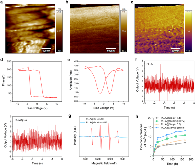
Within the PFM mode of atomic power microscopy (AFM), a tip bias of 10 V was utilized to conduct localized point-to-point piezoelectric response assessments on the pattern materials, aimed toward investigating the piezoelectric traits of the fabric’s floor. Determine 2a illustrates the floor of the chosen piezoelectric materials. The experimental outcomes introduced in Fig. 2b-c point out that the magnitude of the piezoelectric response on the fabric’s floor, following the applying of the tip bias, was 115 pm and a part angle of 167°, respectively. This implies that the fabric’s floor displays a excessive piezoelectric response functionality, additional validating the distinctive piezoelectric properties of the PLLA@Ga composite materials on the nanoscale. The attribute hysteresis loop and pronounced butterfly curve noticed within the PLLA@Ga samples in Fig. 2d-e reveal the fabric’s spontaneous polarization potential beneath the affect of switching voltage, indicating favorable electrical habits. Moreover, the fabric continues to display a sturdy piezoelectric impact beneath ultrasonic mechanical forces, showcasing inherent spontaneous polarization traits, thereby defining the fabric’s non-zero remnant polarization or ferroelectric properties. These piezoelectric and polarization traits present vital proof for the fabric’s potential functions within the biomedical area, notably in diabetic wound therapeutic.
To additional validate the piezoelectric conversion functionality of the ready materials, we examined its output voltage response beneath ultrasonic stress. Determine 2f exhibits that each PLLA and PLLA@Ga exhibit distinct output voltage alerts when subjected to dynamic US stress, indicating that the fabric can successfully convert US power into electrical power, additional confirming its glorious piezoelectric efficiency. Along with the piezoelectric response assessments, we additionally investigated the flexibility of PLLA@Ga to generate ROS beneath US stimulation. For this function, we employed electron spin resonance (ESR) spectroscopy to evaluate the technology of hydroxyl radicals (•OH) beneath US stimulation. As proven in Fig. 2g, a distinguished •OH attribute peak was noticed within the ESR spectrum, indicating that the PLLA@Ga composite materials can successfully produce hydroxyl radicals beneath US stimulation. This implies that PLLA@Ga bear particular reactions beneath US stimulation, changing hydrogen peroxide (H2O2) into extremely reactive •OH radicals, thereby additional enhancing the fabric’s antibacterial properties and selling wound therapeutic.
In abstract, the experimental outcomes point out that the PLLA@Ga composite materials not solely possesses glorious piezoelectric efficiency but in addition enhances antibacterial results by way of ROS technology beneath US stimulation. This broadens the applying prospects of the fabric in diabetic wound therapeutic, notably in modulating immune responses, selling wound restore, and stopping infections.
On this research, we immersed PLLA@Ga composites in PBS buffer resolution (pH = 7.4) and recorded the focus modifications of Ga3+ within the resolution at totally different time factors, each within the presence and absence of US, as illustrated in Fig. 2h. Over the interval from 6 h to 7 days, the focus of gallium ions progressively elevated, indicating that the PLLA@Ga materials underwent an ion launch course of within the PBS resolution. In comparative experiments, we noticed that the Ga3+ launch charge within the US (+) group was considerably greater than that within the US (-) group, with the whole quantity of Ga3+ on the finish of the experiment additionally markedly larger within the US (+) group. This implies that the applying of ultrasound can successfully speed up the discharge of Ga3+ from PLLA@Ga composites, seemingly resulting from mechanical results induced by ultrasound that improve the dissolution or diffusion charge of the ions. Moreover, we investigated the impression of pH on Ga3+ launch. Experimental outcomes indicated that beneath decrease pH situations (pH = 5.5), the discharge charge of Ga3+ from PLLA@Ga was considerably accelerated. This phenomenon could also be associated to the elevated degradation charge of the fabric in an acidic surroundings, facilitating the leaching of Ga3+ from the composite. On condition that wound websites typically exhibit localized acidic situations, notably in the course of the inflammatory part or within the presence of bacterial an infection, decrease pH values could promote the discharge of extra Ga3+, thereby enhancing the antibacterial efficacy of the fabric.
Characterization of PLLA@Ga. (a) PFM picture of PLLA@Ga materials floor. (b) Piezoelectric amplitude of PLLA@Ga. (c) Part angle of PLLA@Ga. (d) Butterfly cycle curve of PLLA@Ga. (e) Piezoelectric flip curve of PLLA@Ga. (f) The output voltage of PLLA@Ga beneath US stimulation. (g) ESR of PLLA@Ga with US or with out US. (h) Ga3+ focus was decided by ICP-OES after immersion of PLLA@Ga in PBS at totally different pH (pH 7.4 and pH 5.5) for various occasions with or with out US
In vitro results of PLLA@Ga + US
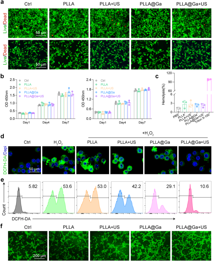
The significance of excellent biocompatibility can’t be overstated within the scientific software of biomaterials. To evaluate this, we performed in vitro biocompatibility testing on numerous supplies utilizing stay/lifeless staining, CCK8, and hemolysis assays. Initially, HUVEC and RS1 cells have been cultured on totally different spinnerets with or with out ultrasound stimulation. Following a 24-hour incubation interval, stay/lifeless staining was carried out. The outcomes depicted in Fig. 3a illustrate that after the remedies, the vast majority of HUVEC and RS1 cells exhibited excessive viability (inexperienced fluorescence) with minimal cell dying (pink fluorescence). These findings counsel that PLLA@Ga demonstrates biocompatibility and helps cell survival. The impression of PLLA@Ga on cell proliferation was additional examined by way of CCK8 evaluation. The CCK-8 assay outcomes demonstrated that cell proliferation elevated over the course of 1, 4, and seven days of tradition, indicating that PLLA@Ga helps cell progress with out cytotoxicity (Fig. 3b). In contrast to the 1-day tradition interval, cell absorbance values elevated after 4 and seven days of tradition with all remedies, suggesting that PLLA@Ga successfully enhances cell proliferation. Hemolysis experiments additionally confirmed the blood compatibility of PLLA@Ga, as not one of the biomaterial remedies resulted in vital hemolysis (Fig. 3c). These findings spotlight the favorable biocompatibility, non-hemolytic nature, and help for cell adhesion, progress, and proliferation offered by PLLA@Ga composite electrospinning.
The standard traits of diabetic wounds embrace elevated ranges of ROS, indicating that the wound is in a state of persistent oxidative stress. Immunofluorescence staining utilizing DCFH-DA was employed to evaluate the flexibility of macrophages within the oxidative stress harm remedy group to clear intracellular. The management group uncovered to H2O2 confirmed greater DCFH-DA staining, suggesting the induction of oxidative stress. Notably, after remedy with PLLA@Ga + US, the fluorescence sign decreased to ranges much like these of cells not uncovered to H2O2 (Fig. 3d). The outcomes are additional supported by movement cytometry outcomes (Fig. 3e). These findings counsel that PLLA@Ga + US can improve the antioxidant capability of macrophages by scavenging inside oxidants. Enhancing angiogenesis is essential for tissue restore. Due to this fact, we employed the HUVEC tube formation assay to guage the impression of PLLA@Ga + US remedy on angiogenesis. The experimental outcomes demonstrated a major improve within the tubular community within the PLLA@Ga + US group in comparison with the management group (as proven in Fig. 3f), indicating that this remedy successfully promotes endothelial cell tube formation and the angiogenic course of. The improved tube formation functionality serves as an indicator of endothelial cell migration, proliferation, and differentiation throughout angiogenesis, offering strong proof for vascular reconstruction within the wound therapeutic course of. Moreover, to validate the angiogenic potential of PLLA@Ga + US, we measured the secretion ranges of vascular endothelial progress issue (VEGF) throughout totally different remedy teams utilizing Elisa (Determine S2). VEGF is a vital issue that promotes angiogenesis by stimulating endothelial cell proliferation and the formation of recent blood vessels. The outcomes revealed that the secretion ranges of VEGF within the PLLA@Ga + US group have been considerably greater than these within the management group, additional substantiating the efficacy of this remedy in selling angiogenesis.
Results of PLLA@Ga + US in vitro. (a) Photographs of stay/lifeless assays of HUVECs (higher panel) and RS-1 cells (decrease panel). (b) CCK-8 assay of HUVECs (left panel) and RS-1 cells (proper panel). (c) Hemolysis assay outcomes of vatious supplies publicity. (d) Fluorescence imaging was employed to evaluate the discount of intracellular ROS in H2O2-stimulated Raw264.7 cells. (e) Evaluation of intracellular ROS in H2O2-stimulated Raw264.7 cells after totally different remedies through movement cytometry. (f) Tube formation through coculturing HUVECs with numerous hydrogels
The noticed improve in cell proliferation and excessive cell viability on PLLA@Ga membranes is in step with earlier research demonstrating the biocompatibility of Ga-doped supplies. For example, Li et al. reported that Ga3+ improve cell adhesion and proliferation by modulating the extracellular matrix surroundings [38]. Moreover, the improved hydrophilicity of PLLA@Ga, as evidenced by the lowered water contact angle, aligns with findings from Solar et al., who highlighted that hydrophilic surfaces promote cell attachment and progress [39]. The antioxidant capability of PLLA@Ga, as demonstrated by the discount in intracellular ROS ranges, can also be supported by research displaying that Ga3+ can scavenge ROS and mitigate oxidative stress [40]. These outcomes collectively counsel that PLLA@Ga not solely helps cell proliferation but in addition creates a positive microenvironment for tissue regeneration.” The improved tube formation noticed within the HUVEC assay signifies that PLLA@Ga promotes angiogenesis, a crucial think about diabetic wound therapeutic. This discovering is especially vital given the impaired angiogenesis sometimes related to diabetic wounds.
Antibacterial efficacy of PLLA@Ga + US
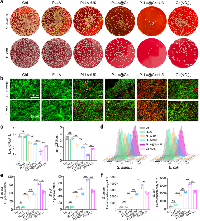
Dressings possessing potent antimicrobial properties are efficient in safeguarding towards pathogenic bacterial contamination, a crucial issue influencing the development of wound therapeutic. The antibacterial effectiveness of PLLA@Ga + US electrospun fibers was assessed by testing them towards numerous micro organism. The bacterial colonies have been quantified publish numerous remedies utilizing SPM. As depicted in Fig. 4a and c, and S1, the bacterial colony rely considerably decreased following the PLLA@Ga + US remedy. The micro organism subjected to varied remedies have been stained utilizing stay/lifeless staining and examined beneath a fluorescence microscope. The findings illustrated in Fig. 4b reveal a considerable improve in bacterial mortality (pink fluorescence) inside the PLLA@Ga + US group compared to different remedies, accompanied by a notable lower within the variety of surviving micro organism (inexperienced fluorescence). These outcomes underscore the potent antibacterial effectiveness of PLLA@Ga + US.
Subsequently, PI staining was employed to evaluate the bactericidal functionality of PLLA@Ga + US (Fig. 4d). For S. aureus, the PI fluorescence depth of the PLLA@Ga + US group elevated by 9.45-fold, 9.13-fold, 1.26-fold, 0.76-fold, and 0.64-fold in comparison with the management group, PLLA group, PLLA + US group, PLLA@Ga group, and Ga(NO3)3 group (Fig. 4e), respectively. Equally, for E. coli, the PI fluorescence depth of the PLLA@Ga + US group elevated by 9.89-fold, 9.13-fold, 1.41-fold, 0.87-fold, and 0.72-fold relative to the management group, PLLA group, PLLA + US group, PLLA@Ga group, and Ga(NO3)3 group (Fig. 4e), respectively. This means that the PLLA@Ga + US remedy can considerably disrupt bacterial cell membranes, enhancing their permeability. Moreover, quantitative evaluation of PI fluorescence depth utilizing a microplate reader corroborated the outcomes obtained from the aforementioned movement cytometry (Fig. 4f). The outcomes confirmed a major correlation between PLLA@Ga + US remedy and elevated PI fluorescence depth, highlighting its potential as an efficient antibacterial technique.
In vitro antibacterial exercise of PLLA@Ga + US. (a) Consultant plates of micro organism colonies after remedy with totally different samples. (b) The fluorescence pictures of stay (inexperienced) and lifeless bacterial (pink). (c) Outcomes of micro organism handled with numerous supplies that shaped colonies after 24 h. (d-e) Consultant movement cytometry plots and quantitative evaluation of PI. (f) Detection of membrane harm in micro organism by way of monitoring PI inflow. Knowledge are introduced as imply ± SD (n = 3). “ns” signify “no vital”; *P < 0.05; **P < 0.01; ***P < 0.001
Antibacterial efficacy of PLLA@Ga + US
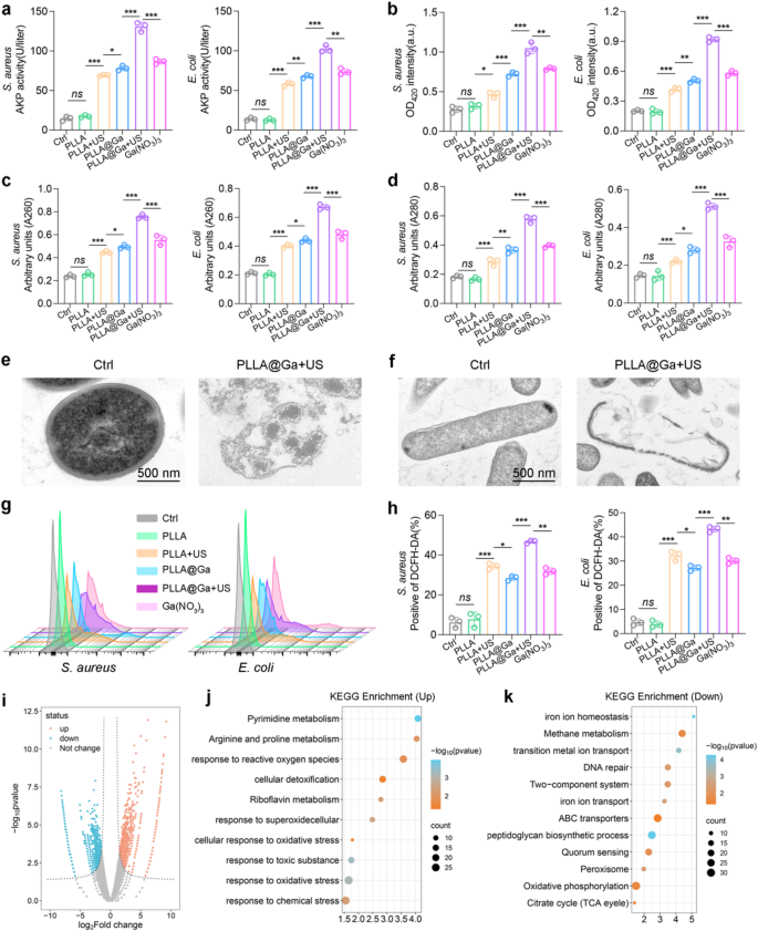
Moreover, the antibacterial impact of PLLA@Ga + US was additional investigated by assessing the extracellular AKP content material and ONPG hydrolysis assay. Following remedy with PLLA@Ga + US, there was a major improve in extracellular AKP focus (Fig. 5a), indicating harm to the bacterial cell wall. This discovery means that this remedy is able to successfully disrupting the bacterial cell wall. ONPG enters the cytoplasm and is subsequently hydrolyzed by cytoplasmic β-galactosidase, indicating a rise in bacterial cell membrane permeability. Notably, the absorbance at 420 nm considerably elevated following PLLA@Ga + US remedy (Fig. 5b), suggesting enhanced bacterial cell membrane permeability. In comparison with different teams, micro organism handled with PLLA@Ga + US exhibited a major improve in nucleic acid and protein absorbance at 260 nm and 280 nm (Fig. 5c-d). These outcomes display that PLLA@Ga + US remedy can considerably alter bacterial membrane permeability, resulting in cytoplasmic leakage. Moreover, TEM outcomes revealed that micro organism handled with PLLA@Ga + US confirmed contracted, blurred, and corrugated cell membranes, whereas the management group displayed {smooth}, intact, and well-defined cell membranes (Fig. 5e).
Subsequently, the potential mechanism of PLLA@Ga + US sterilization was investigated. In keeping with earlier research, piezoelectric supplies can produce a considerable amount of ROS beneath ultrasonic stimulation. Due to this fact, we speculate that this remedy could exhibit antibacterial properties by way of ROS exercise. As proven in Figs. 5g–h, micro organism handled with PLLA@Ga + US exhibited a major improve in ROS manufacturing. Due to this fact, ROS performs an integral function within the mechanism of PLLA@Ga + US sterilization. Moreover, as per earlier studies, Ga3+ possesses antibacterial exercise primarily as a result of micro organism take up Ga and bind it to iron-dependent enzymes [41, 42]. Nonetheless, Ga3+ is unable to be lowered beneath physiological situations, rendering Ga-substituted enzymes unable to hold out the duties of their iron-dependent counterparts, that are important for bacterial progress. Since micro organism can not differentiate between the 2, they’re unable to scale back their consumption of Ga3+, as doing so would additionally lower their consumption of iron. Due to this fact, the potent bactericidal impact of PLLA@Ga + US might be attributed to the numerous quantity of Ga3+ disrupting bacterial iron metabolism and in the end resulting in bacterial dying.
Subsequent, RNA sequencing evaluation was performed on surviving S. aureus to determine gene expression variations between the management group and the PLLA@Ga-treated group beneath US remedy. Volcano plots have been produced to visually signify the differentially expressed genes noticed between the Ctrl group and the PLLA@Ga + US group (Fig. 5i). Make the most of the KEGG enrichment technique to research differentially expressed genes. Within the PLLA@Ga + US group, genes related to ROS, superoxide, and oxidative stress have been considerably upregulated (Fig. 5j), indicating that PLLA@Ga + US remedy can improve oxidative stress in S. aureus. Earlier research have proven that Ga3+ can bind to bacterial cytochromes, inhibiting the electron switch of cytochrome-bound oxygen, resulting in ROS manufacturing and inducing bacterial dying [43]. Due to this fact, PLLA@Ga + US could inhibit bacterial progress by enhancing bacterial oxidative stress. As well as, pathways associated to DNA restore are considerably downregulated within the PLLA@Ga + US group (Fig. 5ok). This implies that DNA restore is successfully inhibited, catalytic reactions inside the micro organism are disrupted, and bacterial morphological regulation is suppressed. Genes associated to iron ion transport have been considerably downregulated within the PLLA@Ga + US group (Fig. 5ok), which might be defined by the micro organism’s lack of ability to tell apart between Ga3+ and Fe3+, ensuing within the uptake of a considerable amount of Ga3+ [44]. Iron is without doubt one of the most vital vitamins for micro organism, concerned in important intracellular redox and electron switch processes. Consequently, PLLA@Ga + US can inhibit bacterial progress by interfering with S. aureus’s iron metabolism.
These outcomes point out that PLLA@Ga + US can inhibit the iron ion transport of S. aureus, scale back the exercise of bacterial antioxidant enzymes, and amplify the oxidative stress of micro organism, thereby enhancing the antibacterial exercise of PLLA@Ga in the course of the SDT course of.
In vitro antibacterial exercise of PLLA@Ga + US. (a) The sensitivity to alkaline phosphatase exercise. (b) The ONPG hydrolysis exercise. (c-d) Leakage of cell contents after publicity to varied remedies was investigated by monitoring the absorbance of extracellular materials at 260 nm (A260) and 280 nm (A280) akin to nucleic acids and proteins, respectively. (e-f) Morphology of micro organism from totally different remedy teams as noticed by TEM. (g) Evaluation of ROS technology was performed utilizing DCFH-DA fluorescent probes by FCM. (h) Evaluation of ROS technology was performed utilizing DCFH-DA fluorescent probes by microplate reader. (i) Volcano map of differentially expressed genes (DEGs). (j-k) KEGG pathway enrichment evaluation. Knowledge are introduced as imply ± SD (n = 3). “ns” signify “no vital”; *P < 0.05; **P < 0.01; ***P < 0.001
Macrophage habits
Quite a few analysis findings point out that the effectivity of wound therapeutic is carefully tied to the native inflammatory response inside the tissue [45,46,47]. Undoubtedly, the immune system considerably influences this intricate course of. Overactive or dysregulated immune responses can worsen tissue harm and impede the therapeutic course of. Therefore, optimum wound therapeutic and restoration transpire inside a harmonious and well-regulated immune surroundings. Macrophages are acknowledged for his or her essential involvement within the immune system and have the flexibility to rework into two distinct phenotypes, M1 (pro-inflammatory subtype) and M2 (pro-repair subtype), relying on the stimuli they encounter. M1 macrophages encourage the event of an inflammatory microenvironment by way of the secretion of varied pro-inflammatory cytokines, together with tumor necrosis factor-α (TNF-α), IL-6, and IL-1β. Conversely, M2 macrophages facilitate tissue restore by releasing cytokines like IL-4, CD206, and IL-10 to determine an anti-inflammatory setting [48, 49]. The importance of this polarization is paramount, as the suitable polarization of M1 and M2 macrophages enormously improves the tissue restore course of. Conversely, extreme activation and polarization can worsen irritation and hinder environment friendly wound therapeutic [50]. Undoubtedly, the host immune response is a vital issue within the wound therapeutic course of facilitated by biomaterials. Given the crucial nature of those immune responses, our focus now shifts to investigating the immunomodulatory impacts of PLLA@Ga + US and its potential to reinforce wound therapeutic by way of modulation of macrophage actions.
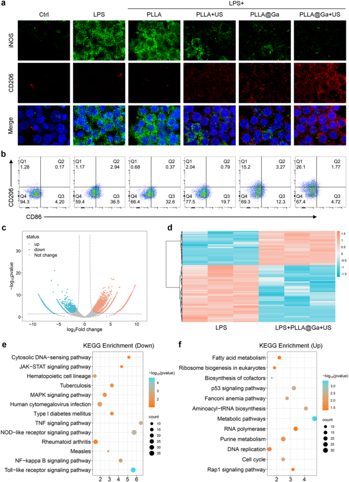
Macrophages have been seeded on totally different fibrous membranes for twenty-four h beneath LPS-induced irritation. The immunomodulatory impact of PLLA@Ga + US was evaluated by analyzing the expression of macrophage M1 and M2 subtype markers by way of immunofluorescence staining. Outcomes depicted in Fig. 6a revealed that after publicity to LPS, macrophages exhibited elevated ranges of iNOS (inexperienced fluorescence). Conversely, remedy with PLLA + US, PLLA@Ga, and PLLA@Ga + US, notably the latter, considerably decreased iNOS expression, suggesting an inhibitory impact on its expression (Determine S3a). Moreover, the fluorescence depth of CD206 notably elevated following remedies with PLLA + US, PLLA@Ga, and PLLA@Ga + US in comparison with LPS remedy. Notably, PLLA@Ga + US exhibited essentially the most pronounced impact on CD206 fluorescence depth (Determine S3b). These findings point out that PLLA@Ga + US has the potential to counteract the LPS-induced inflammatory microenvironment, suppressing macrophage M1 polarization whereas selling M2 polarization.
The impression of various remedies on macrophage polarization was subsequently evaluated by way of movement cytometry evaluation. The outcomes revealed a major lower within the proportion of CD86-positive cells (M1 marker) and a notable improve in CD206-positive cells (M2 marker) following remedy with PLLA + US, PLLA@Ga, and PLLA@Ga + US, notably PLLA@Ga + US. Particularly, the distribution of CD86-positive cells within the management, LPS, LPS + PLLA, LPS + PLLA + US, LPS + PLLA@Ga, and LPS + PLLA@Ga + US teams have been 4.20%, 36.5%, 32.6%, 19.7%, and 12.3%, and 4.72%, respectively, whereas the proportions of CD206-positive cells have been 1.28%, 1.17%, 0.68%, 2.04%, 15.2%, and 26.1% (Fig. 6b). These outcomes counsel an in depth relationship between PLLA@Ga + US remedy and the shift of macrophages in the direction of the anti-inflammatory M2 phenotype. The downregulation of CD86 and upregulation of CD206 spotlight the flexibility of PLLA@Ga + US to direct macrophages in the direction of the reparative M2 subtype, which is significant for efficient wound therapeutic in circumstances of an infection.
Moreover, an in-depth investigation into the consequences of PLLA@Ga + US on macrophages was performed utilizing RNA-Seq. Differentially expressed genes (DEGs) have been recognized with a p-value of lower than 0.05 and a fold change (FC) larger than 2. The volcano plot in Fig. 6c vividly illustrates the substantial variety of DEGs between the PLLA@Ga + US remedy group and the LPS group. The heatmap constructed based mostly on these differential genes highlights the modifications in gene expression between the LPS group and the LPS + PLLA@Ga + US group (Fig. 6d). KEGG pathway enrichment evaluation additional revealed particular signaling pathways related to macrophage phenotype transformation (Fig. 6e-f). The evaluation indicated that after PLLA@Ga + US remedy, inflammatory pathways associated to M1 macrophage polarization, together with MAPK signaling pathway, TOR-like receptor signaling pathway, NF-kappa B signaling pathway, and Nod-like signaling pathway, have been considerably inhibited. As well as, upregulation of anti-inflammatory pathways was noticed, notably these related to M2 macrophage polarization, such because the TGF-β signaling pathways. This alteration in gene expression means that macrophages could also be reprogrammed in the direction of a extra reparative phenotype, thereby selling tissue regeneration.
To validate the RNA-Seq findings, the impression of PLLA@Ga + US remedy on the expression of inflammation-related genes in macrophages was examined utilizing qPCR. After 24 h of tradition, the expression ranges of typical M1 macrophage markers (together with IL-1β, TNF-α, and iNOS) within the PLLA@Ga + US remedy group have been discovered to be considerably decrease in comparison with these within the LPS remedy group alone (Determine S1c). Conversely, the expression degree of the M2 macrophage marker IL-10 confirmed a major improve, which aligns with the RNA-Seq outcomes. Elisa was employed to measure the secretion of inflammatory elements. The PLLA@Ga + US remedy demonstrated a major lower within the secretion ranges of pro-inflammatory cytokines like IL-1β and TNF-α (Determine S1d). These findings present extra proof supporting the flexibility of PLLA@Ga + US remedy to modulate macrophage transformation in the direction of the anti-inflammatory subtype. These findings spotlight the therapeutic potential of PLLA@Ga + US in modulating macrophage behaviour, thereby influencing the general inflammatory response and selling a healing-favourable microenvironment.
The traits of macrophages are influenced by numerous elements comparable to their inducers, properties, and floor markers. This analysis concerned the creation of electrospun PLLA@Ga with piezoelectric capabilities to research its impression on macrophage phenotype. The findings indicated that making use of PLLA@Ga with ultrasonic stimulation resulted in a discount of M1-type macrophages induced by LPS, whereas selling a rise in M2-type macrophages. By evaluating with ICP outcomes, we discovered that the focus of Ga ions in PLLA@Ga is 11.74 mg/L. Earlier research have demonstrated that free Ga ions can inhibit the expression of inflammatory genes [51], indicating that Ga ions play an indispensable function within the anti-inflammatory results of PLLA@Ga. Moreover, a number of analysis has proven that the piezoelectric impact induced by mild US additionally possesses sure anti-inflammatory properties [30], which has been additional confirmed on this research.
Research on the in vitro immunomodulatory of PLLA@Ga + US. (a) Expression of iNOS and CD206 following totally different hydrogel remedies. (b) Evaluation of CD86 and CD206 in macrophages following numerous remedies through movement cytometry. (c) Volcano map of differentially expressed genes (DEGs). (d) Warmth map of differentially expressed genes (DEGs). (e) KEGG enrichment for the down-regulated pathways. (f) KEGG enrichment for the up-regulated pathways
In vivo wound therapeutic of PLLA@Ga + US
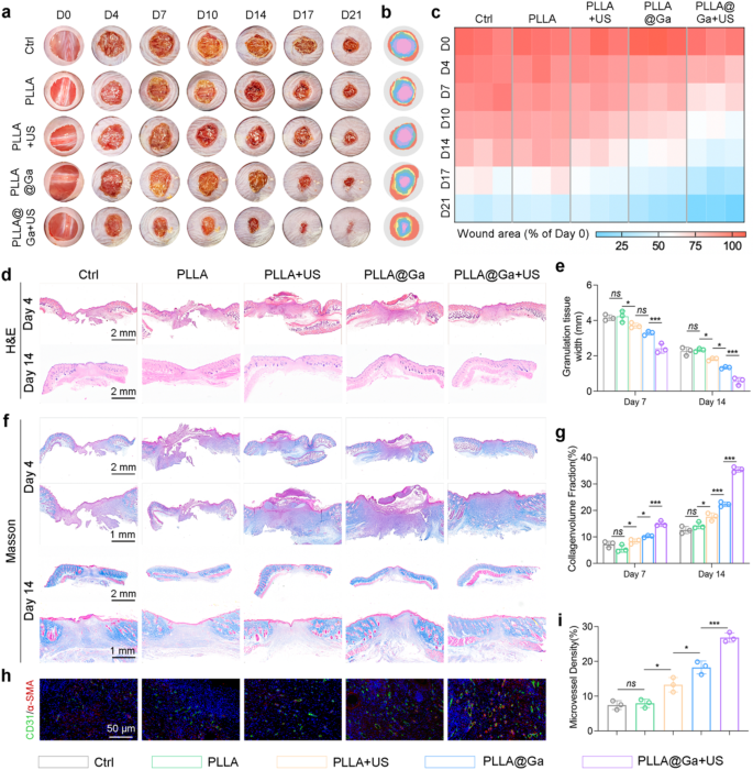
Within the context of diabetes-related wounds, extended delays in therapeutic pose a heightened danger of bacterial an infection, thereby impairing the physique’s immune response and impeding the wound restoration course of. This situation can probably escalate to extreme infections and significant well being issues [52, 53]. Therefore, expediting the wound therapeutic course of is essential for each stopping and managing infections. This analysis delves into exploring the impression of PLLA@Ga on wound therapeutic inside an contaminated diabetic mouse mannequin. Mice have been induced with diabetes by injecting STZ and creating a ten mm full-thickness wound on mice with blood glucose ranges exceeding 16.65 mmol/L, adopted by contamination with micro organism. Wounds handled with saline (management group), PLLA, PLLA + US, PLLA@Ga, and PLLA@Ga + US have been photographed and evaluated on days 0, 4, 7, 10, 14, 17, and 21. As proven in Fig. 7a-b, wounds handled with PLLA@Ga + US healed sooner in comparison with different remedy teams. The therapeutic charges of wounds handled with PLLA@Ga + US on days 4, 7, 10, 14, 17, and 21 have been 21.5%, 36.9%, 48.2%, 57.3.73%, and 93.7% respectively, whereas the management group solely achieved a therapeutic charge of 65.2% on day 21 (Fig. 7c).
On the seventh and twenty first day post-operation, histological examination of the contaminated wound pores and skin was performed utilizing H&E and Masson’s trichrome staining for tissue evaluation. Quantitative evaluation of granulation tissue size, as proven in Fig. 8d-e, indicated a considerably accelerated wound contraction charge within the PLLA@Ga + US remedy group in comparison with the others. Ample collagen deposition is essential for tissue power and therapeutic in the course of the transforming course of. We employed Masson’s trichrome staining to guage new collagen. By day 21, the management group nonetheless had residual scabs, indicating delayed therapeutic (Fig. 8f). In distinction, wounds handled with PLLA@Ga + US exhibited a transparent pink epidermal layer and extra collagen. Moreover, PLLA@Ga + US remedy additionally enhanced persistent wound therapeutic, selling efficient regeneration of dermal appendages (Fig. 8g). In conclusion, the research findings counsel that PLLA@Ga + US remedy enhances the therapeutic charge of infection-related diabetic wounds in comparison with different teams. Analysis findings point out that profitable wound therapeutic depends on angiogenesis to offer essential vitamins and oxygen. Thus, enough angiogenesis performs a significant function within the wound therapeutic course of [54]. The research assessed the presence of neoangiogenic markers, particularly cell adhesion molecule 1 (CD31) and α-smooth muscle actin (α-SMA), in sections of mouse pores and skin wounds (Fig. 8h-i). The outcomes revealed notably elevated ranges of CD31 and α-SMA in wounds handled with PLLA@Ga + US, underscoring their noteworthy potential for selling wound therapeutic.
PLLA@Ga + US promotes diabetic wound therapeutic. (a-b) Images of wounds at predetermined time factors and superimposed pictures of wounds after numerous remedies. (c) Areas of unhealed wounds in every group at totally different time intervals. (d) H&E staining. (e) Corresponding quantitative evaluation of the granulation tissue width. (f) Masson’s trichrome staining. (g) Statistical evaluation of collagen deposition in the course of the remolding part. (h) Consultant immunofluorescence staining of microvessel of wound. (i) Statistical evaluation of microvessel density. Knowledge are introduced as imply ± SD (n = 3). “ns” signify “no vital”; *P < 0.05; ***P < 0.001
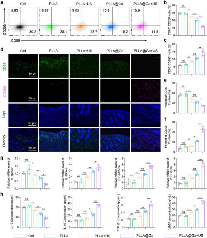
The efficient remedy for diabetic contaminated wounds entails not solely quickly eradicating micro organism but in addition reversing the inflammatory response within the diabetic microenvironment. Histological evaluation signifies that PLLA@Ga + US remedy can obtain speedy therapeutic of diabetic contaminated wounds. In vitro experiments display that PLLA@Ga + US remedy can inhibit macrophage M1 polarization and promote M2 polarization. Due to this fact, it’s affordable to take a position that PLLA@Ga + US remedy can reverse the inflammatory microenvironment of diabetic wounds in vivo, thereby selling wound therapeutic. Accumulate wounds subjected to totally different remedies and analyze the single-cell suspensions utilizing movement cytometry to quantify macrophage populations. Determine 8a-c display that, in comparison with different remedy teams, the PLLA@Ga + US remedy group exhibited fewer M1-like macrophages (CD86+CD206−) and a major improve in M2-like macrophages (CD86−CD206+). Immunofluorescence staining of tissue-associated M1 (CD86) and M2 (CD206) macrophages revealed that PLLA@Ga + US remedy considerably enhanced M2 macrophage markers whereas lowering the expression of M1 macrophage markers (Fig. 8d-f). These findings affirm that PLLA@Ga + US remedy improves the native immune microenvironment of diabetic contaminated wounds by lowering CD86+ M1 macrophages and rising CD206+ M2 macrophages close to the wound, which is extra conducive to wound therapeutic. Furthermore, PCR and Elisa have been employed to quantify the gene expression and protein secretion ranges of M1 cell elements (associated to the pro-inflammatory marker IL1β) and M2 cell elements (anti-inflammatory and angiogenesis markers IL10, TGFβ1, and VEGF) (Fig. 8g-h). The evaluation outcomes revealed a major lower in M1 macrophages issue expression ranges and a rise in M2 macrophages issue protein ranges within the PLLA@Ga + US remedy group, surpassing different remedy teams. These findings point out that PLLA@Ga + US can successfully promote the therapeutic of diabetic contaminated wounds.
Macrophages play a vital function within the immune response to tissue harm. In diabetic wound infections, the transition of macrophages from an inflammatory (M1) to an anti-inflammatory (M2) state is disrupted, impeding wound therapeutic [55, 56]. Our in vitro and in vivo research display that PLLA@Ga + US successfully reduces the variety of M1 macrophages whereas rising the variety of M2 macrophages, thereby enhancing the expression of anti-inflammatory and angiogenic cytokines, together with IL-10, VEGF, and TGF-β1. Particularly, IL-10 is a cytokine that alleviates irritation and inhibits oxidative processes. Vascular endothelial progress issue performs a crucial function in recruiting endothelial cells, thereby initiating angiogenesis. Moreover, TGF-β1 recruits endothelial cells and fibroblasts to the wound web site, selling the synthesis of ECM [57]. In conclusion, PLLA@Ga + US exhibits vital potential in accelerating the therapeutic of diabetic-infected wounds. By successfully modulating macrophage phenotypes, PLLA@Ga + US aids in suppressing irritation response, selling angiogenesis, and tissue transforming.
Affect of the PLLA@Ga + US on macrophage polarization in vivo. (a-c) Stream cytometry of M1and M2 macrophages retrieved from the wound tissue. (d) Immunofluorescence staining of tissue sections on the wound web site for CD86 and CD206. (e-f) Statistical evaluation regarding the proportion of CD86 (f) and CD206 (g) optimistic macrophages. (g) Gene expression ranges of IL-1β, IL-10, VEGF, and TGF-β1 in wound tissues. (h) Secretion of IL-1β, IL-10, VEGF, and TGF-β1 in wound tissues assessed by Elisa. Knowledge are introduced as imply ± SD (n = 3). “ns” signify “no vital”; *P < 0.05; **P < 0.01; ***P < 0.001
On this research, we evaluated the consequences of PLLA + US remedy on main organs, together with the center, liver, spleen, lungs, and kidneys, by way of H&E staining. The outcomes indicated that none of those organs exhibited vital poisonous responses following PLLA@Ga + US remedy (Determine S4). Particularly, the staining pictures didn’t reveal any tissue harm, irritation, or different poisonous traits. The tissue structure of the center, liver, spleen, lungs, and kidneys remained intact, with regular mobile morphology and no evident mobile infiltration or tissue destruction. Moreover, the blood biochemical parameters of mice, comparable to whole protein (TP), albumin (ALB), globulin (GLO), whole bilirubin (TBIL), alanine aminotransferase (ALT), aspartate aminotransferase (AST), urea, and glucose (GLU), confirmed no vital modifications after totally different remedies (Determine S5), indicating that PLLA@Ga + US possesses glorious biocompatibility and biosafety. These findings counsel that PLLA@Ga + US remedy demonstrates good biocompatibility with these important organs and doesn’t elicit poisonous responses, thereby offering a security assurance for its scientific software. Moreover, the ultrasound remedy didn’t trigger any harm to the organs, additional validating that the mixed motion of PLLA composites and ultrasound can promote wound therapeutic whereas sustaining systemic security.
Regardless of the promising in vitro and in vivo outcomes, a number of challenges stay for the scientific translation of PLLA@Ga. These embrace the scalability of the electrospinning course of, the long-term stability of the piezoelectric properties, and the potential want for repeated ultrasound remedies in scientific settings. Moreover, the protection and efficacy of PLLA@Ga in human sufferers should be rigorously evaluated by way of scientific trials. You will need to notice that whereas PLLA@Ga exhibits promising outcomes, its efficiency ought to be contextualized inside the broader area of superior wound dressings. For example, silver-based dressings and hydrogels have been broadly used for his or her antibacterial properties, whereas progress factor-loaded scaffolds have proven efficacy in selling angiogenesis [58,59,60]. In comparison with these supplies, PLLA@Ga provides a singular mixture of piezoelectricity and Ga ion launch, which can present synergistic advantages in modulating the immune microenvironment and selling tissue restore. Nonetheless, additional research are wanted to straight examine the efficacy of PLLA@Ga with these present supplies.

