Preparation and characterization of PEG-GVs
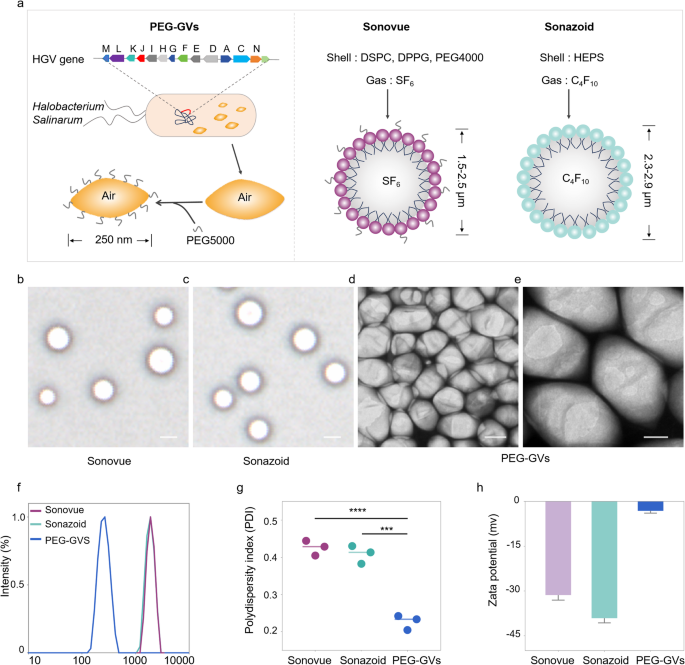
Not like Sonovue and Sonazoid produced via chemical synthesis with lipid parts and inert gases like sulfurhexafluoride (SF6) or perfluorobutane (C4F10), GVs are synthesized by way of a organic synthesis route. Usually, GVs are encoded by gvp gene clusters within the genome and assembled into gas-filled protein shells together with the expansion of microorganisms. Firstly, we extracted GVs from Halobacterium Salinarum via lysis methodology as reported beforehand [24,25,26]. To scale back their immunogenicity to physique, PEG5000 was subsequently conjugated onto the floor of GVs via amidation response within the presence of EDC and NHS, ensuing within the formation of PEG-GVs (Fig. 2a, left panel). The construction and composition of PEG-GVs additionally diverge considerably from Sonovue and Sonazoid. PEG-GVs function gvp protein shells and air core. In distinction, Sonovue consists of DSPC/DPPG shells encapsulating an SF6 core, and Sonazoid predominantly consists of hydrogenated egg phosphatidyl serine (HEPS) shells with a C4F10 core (Fig. 2a, proper panel) [27]. This structural disparity can also be mirrored of their morphological look. Below the microscope, Sonovue and Sonazoid each exhibit a spherical form, whereas PEG-GVs show an ovary form below the scanning electron microscope (SEM) (Fig. 2b-2e). The hydrodynamic measurement of the UCAs was additionally measured by way of dynamic gentle scattering (DLS). Notably, GVs and PEG-GVs are considerably smaller, with a imply hydrodynamic diameter of 213.60 ± 1.71 nm, 252.90 ± 3.08 nm, respectively (Determine S1), versus Sonovue and Sonazoid which have particle sizes within the micrometer vary: 2.18 ± 0.78 μm and a pair of.36 ± 0.95 μm, respectively (Fig. 2f). The polydispersity index (PDI) additional differentiates these brokers. The worth of this index for PEG-GVs is 0.23 ± 0.02, which is considerably decrease than that of Sonovue 0.43 ± 0.02 and Sonazoid 0.41 ± 0.02 (Fig. 2g). In our in vitro statement experiments, we additionally discovered that in comparison with Sonovue and Sonazoid, PEG-GVs exhibit superior dispersion traits and long-term stability (Determine S2). The zeta potentials of PEG-GVs, Sonovue, and Sonazoid are measured at -3.17 ± 0.78 mV, -31.38 ± 1.69 mV, and − 39.16 ± 1.55 mV, respectively (Fig. 2h). Thus, our outcomes reveal that PEG-GVs have uniform nanoscale particle measurement and excellent stability.
Preparation and characterization of PEG-GVs. (a) Composition and construction diagram of biosynthetic PEG-GVs, Sonovue and Sonazoid. b–c, the morphologies of Sonovue (b) and Sonazoid (c) noticed below microscope. Scale bar: 2.5 μm. d–e, SEM pictures of PEG-GVs. Scale bar: 100 nm (d), 50 nm (e). f–h, Particle measurement distribution (f), PDI (g) and Zeta potential (h) of PEG-GVs, Sonovue and Sonazoid. *** p < 0.001, **** p < 0.0001
Acoustic traits and in vitro imaging of PEG-GVs
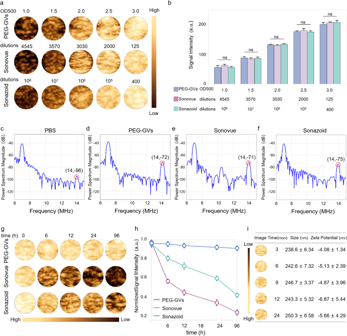
To facilitate comparability of PEG-GVs with Sonovue and Sonazoid in subsequent experiments, we first optimized the concentrations of PEG-GVs to attain a comparable imaging depth to Sonovue and Sonazoid. As depicted in Fig. 3a-3b, we are able to see that the CEUS imaging intensities of PEG-GVs at OD500 1.0, 1.5, 2.0, 2.5 and three.0 had been comparable with Sonovue at 4545-, 3570-, 3030-, 2000-, 125-fold dilutions and Sonazoid at 108-, 107-, 106-, 105-, 400-fold dilutions, respectively. Usually, nonlinear imaging modes rely upon harmonic indicators from microbubble or nanobubble oscillations excited by ultrasound waves, bettering distinction specificity of CEUS. Particularly, the second harmonic indicators are essential for CEUS imaging. Subsequently, we in contrast the harmonic sign traits of PEG-GVs, Sonovue and Sonazoid. Transmitting at 7 MHz, we noticed substantial second-harmonic indicators in PEG-GVs at 14 MHz in comparison with PBS management. As anticipated, Sonovue and Sonazoid produced substantial second-harmonic indicators at 14 MHz totally different from Sonazoid and PEG-GVs, Sonovue additionally produced ultraharmonic indicators (3/2 f0) at 10.5 MHz (Fig. 3e). This phenomenon primarily arises from versatile and nonlinear deformable phospholipid shells and was additionally noticed by different group [28]. Quantitative evaluation confirmed the second harmonic indicators of Sonovue, Sonazoid and PEG-GVs had 25.85 ± 1.70 dB, 22.57 ± 3.03 dB, and 24.00 ± 1.29 dB larger energy spectrum magnitude than the PBS answer on the transmitted frequency (Determine S3). Notably, PEG-GVs produced comparable nonlinear second-harmonic sign intensities, comparable with the industrial Sonovue, Sonazoid. To match the imaging stability of those UCAs, PEG-GVs (OD500 3.0) and Sonovue and Sonazoid (at corresponding concentrations with comparable CEUS imaging intensities) had been saved at 4 °C and imaged at totally different time intervals (Fig. 3g-h). The outcomes confirmed that the distinction imaging depth of Sonovue quickly declined to beneath 50% in a number of hours and beneath 30% after 96 h, whereas that of Sonazoid declined extra slowly but additionally dropped beneath 50% at 96 h. In stark distinction, PEG-GVs confirmed no important sign lower through the 96-h statement interval (Fig. 3h). Moreover, it was additionally noticed that Sonovue and Sonazoid considerably vanished over time, whereas the PEG-GVs largely retained their unique sign depth (Fig. 3g), indicating the wonderful imaging stability of PEG-GVs relative to industrial Sonovue and Sonazoid. To additional assess the long-term stability of PEG-GVs, we prolonged the statement interval to 24 months and detected GVs’ imaging efficiency, particle measurement and zeta potential. We discovered that PEG-GVs nonetheless remained secure in imaging efficiency, particle measurement and zeta potential all through 24-month statement interval (Fig. 3i). These outcomes point out that PEG-GVs saved wonderful CEUS imaging efficiency and strong stability, facilitating their biomedical software and long-term preservation.
Acoustic traits and in vitro imaging of PEG-GVs. a-b, In vitro nonlinear distinction imaging (a) and quantification (b) of PEG-GVs, Sonovue and Sonazoid on the totally different concentrations. c-f, In vitro nonlinear sign spectra of PBS answer (c), PEG-GVs (d), Sonovue (e) and Sonazoid (f). g-h, The distinction pictures (g) and quantification (h) of the three UCAs saved at 4°C for various time from 0 h to 96 h. i, In vitro nonlinear distinction imaging, particle measurement, and zeta potential of PEG-GVs saved at 4°C for 3, 6, 9, 12, 24 months.
In vivo CEUS imaging of PEG-GVs in regular liver tissue
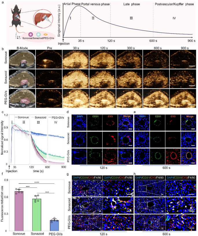
Primarily based on the excellent stability and imaging efficiency of PEG-GVs in vitro, we additional in contrast the liver distinction imaging efficiency of PEG-GVs with Sonovue and Sonazoid in wholesome C57BL/6J mice. The experimental course of and phases of ultrasound distinction agent infusion are depicted in Fig. 4a, displaying the everyday distinction echo sign enhancement modes within the arterial part, portal venous part, late part and postvascular part (additionally known as Kupffer part for Sonazoid). To scale back particular person variations, we intravenously administered these UCAs in the identical mice at 30-min intervals, adopted by a brief high-power burst earlier than subsequent injection. As proven in Fig. 4b, the echo indicators of liver acquired with Sonovue had been enhanced within the arterial part (30 s), subsided within the portal venous part (120 s), and weakened quickly after 300 s. Sonazoid has a chronic imaging time in Kupffer part and gives extra particulars for liver illness analysis. Our outcomes present that the liver echo sign of Sonazoid was considerably enhanced within the arterial part (30 s), barely lowered within the delayed part (300 s), considerably decreased within the Kupffer part (600 s), and turning into weak after 900 s. Apparently, the liver echo with PEG-GVs remained secure throughout all phases, a lot stronger than Sonovue and Sonazoid, particularly within the late and Kupffer phases (Fig. 4b). Quantitative knowledge additional revealed that the imaging depth of Sonovue and Sonazoid declined quickly subsequent to the portal part (120 s), whereas PEG-GVs remained at a excessive degree, with a decline price of lower than 20% in 600 s (Fig. 4f) and nonetheless lower than 30% till 900 s (Fig. 4c). The normalized AUC (Space below curve) 900 s of PEG-GVs was larger than that of Sonazoid and Sonovue, being 2.72- instances of Sonazoid and 1.82 instances of Sonovue, respectively (Determine S4). Clearly, this extended imaging length provides longer time for evaluation, thereby resulting in extra exact analysis and environment friendly interventional operation for medical doctors.
CEUS imaging efficiency and mechanisms of PEG-GVs in regular liver. a, Diagram of regular liver CEUS and its 4 phases when imaging of liver in vivo, scale bar: 5 mm. b, The CEUS pictures of various phases in the identical mice after systemic administration of Sonovue, Sonazoid and PEG-GVs at 30 min intervals. The “L”, “GIT”, “SP” represents liver, abdomen and intestines, and backbone on the B-mode pictures, respectively. c, The normalized sign depth curves of CEUS pictures in Fig. 4b. d–f, Immunofluorescence pictures of vessels (stained with anti-CD31) and CY3-labeled UCAs at 120 s (d) and 600 s (e) after systemic administration. f, The quantification of fluorescence discount price in Fig. 4d. Scale bar: 40 μm. *** p < 0.001, **** p < 0.0001.(n = 5), g–h, Immunofluorescence pictures of liver tissues at 120 s (g) and 600 s (h) after systemic administration. Kupffer cells had been stained with anti-F4/80 and UCAs had been stained by CY3 dye. Scale bar: 40 μm.
To additional elucidate the mechanisms underlying extended imaging length, we carried out immunofluorescence staining utilizing CY3-labeled PEG-GVs and DIO-labeled Sonazoid and Sonovue. Immunofluorescence staining of liver sections revealed a substantial quantity of PEG-GVs penetrated past blood vessels and achieved liver cells. In distinction, scarce Sonazoid and Sonovue may infiltrate blood vessels and settled round blood vessels. Notably, we witnessed the progressive adhesion of PEG-GVs to the vascular partitions after systemic administration. Bubble adhesion was nonetheless observable at 120 s and annular adhesion to the complete blood vessel wall at 600 s after injection (Fig. 4d, 4e, Determine S5). This PEG-GVs’ adherence to the blood vessel partitions might be noticed for as much as 1 h (Determine S6), which could partly clarify their extended imaging length of liver. Contemplating Sonazoid’s distinct benefit within the Kupffer part as UCAs, we additional investigated Kupffer cell uptake of various UCAs in liver tissue. Our findings revealed that Sonazoid and PEG-GVs, however not Sonovue, had been conspicuously internalized by Kupffer cells at 120 s and 600 s. Furthermore, the uptake of PEG-GVs elevated over time (Fig. 4g, 4h). Collectively, our knowledge indicated PEG-GVs possesses excellent CEUS functionality within the liver, which can attribute to their adhesion and penetration past blood vessels and phagocytosis by Kupffer cells.
In vivo CEUS imaging of PEG-GVs in small liver tumor
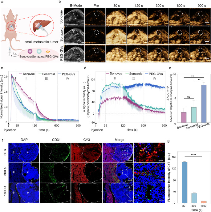
The Kupffer part in liver CEUS has been demonstrated to boost the detection price and specificity of small liver tumor lesions [3, 29]. In gentle of the discovering that Kupffer cells can phagocytose PEG-GVs, we hypothesized that they may play a big position within the analysis of liver tumors. To check it, we employed a murine mannequin of liver metastatic tumors to discover the attainable worth of PEG-GVs in detecting minuscule hepatic lesions (Fig. 5a). After the systemic administration, all of the three UCAs exhibited fast perfusion and fast regression within the tumor (Fig. 5b-5c). Moreover, we noticed a sustained excessive enhancement of PEG-GVs within the regular liver tissue, attributing to their vascular adhesion and Kupffer cell uptake as described above. Apparently, PEG-GVs didn’t exhibit a sustained enhancement in liver tumor tissue attributable to EPR results however regressed quickly. This results in a persistently decrease imaging depth of tumors relative to peripheral liver tissue, particularly within the late part and Kupffer part (Fig. 5d). Quantitative evaluation of tumor-subtracted hepatic parenchyma AUC revealed PEG-GVs had 3.62-fold and a pair of.44-fold larger than Sonovue and Sonazoid, respectively (Fig. 5e). The immunofluorescence outcomes additional confirmed that PEG-GVs didn’t accumulate within the tumor space over time however declined quickly as a substitute, whereas it scarcely decreased within the surrounding regular liver tissue. This is perhaps attributed to the truth that PEG-GVs don’t adhere to the vascular endothelium of tumor tissue, thereby failing to stay within the blood vessels for an prolonged interval (Fig. 5f, Determine S7). Quantitative knowledge additionally demonstrated that PEG-GVs within the tumor decreased to solely 24.3% of peak depth after 300 s of injection and fewer than 10% after 1800 s (Fig. 5g).
CEUS imaging efficiency and mechanism of PEG-GVs in liver tumor. a, Diagram of CEUS of three UCAs in small liver metastatic tumors. b, CEUS pictures of Sonovue, Sonazoid and PEG-GVs at totally different time factors after systemic administration,scale bar: 5mm. c, The normalized sign depth curves of tumors in Figure5b. d-e, CEUS sign depth of peripheral liver tissue (d) and the quantification of AUC distinction between the encircling liver tissue and tumor at 900 s (e). f-g, Blood vessels immunofluorescence staining by anti-CD 31 (f) and quantification (g) of liver metastatic tumors at 30 s, 300 s, 1800 s after CY3-labeled PEG-GVs administration. # and * characterize regular liver and tumor space, respectively. Scale bar: 1000 µm. **** p < 0.0001.
PEG-GVs are nanoscale with roughly 250 nm particle measurement whereas the endothelial house in neovascularized tumors is 380–780 nm, making them simply cross via the tumor vessel by way of the EPR impact [30, 31]. The imaging efficiency of PEG-GVs might assist visualize the EPR course of of those nanobubbles by ultrasound in an actual time method. In the meantime, the fast regression of PEG-GVs from liver tumor additionally challenges the standard idea that the EPR impact inflicting from tumor leaky vessel doesn’t essentially delay the retention time of nanoparticles in a tumor, a minimum of for nanobubbles resembling PEG-GVs. This distinctive phenomenon additionally renders it possible for PEG-GVs to precisely outline tumor boundaries, thereby facilitating early analysis. As for the mechanisms, the attainable causes is perhaps attributed to the excessive permeability of tumor blood vessels, which endows these nanobubbles with larger penetrating functionality into the tumor, however in the meantime it might additionally facilitate their simpler washout from the tumor in comparison with the conventional liver tissue. The structural defect of glycocalyx on the floor of tumor blood vessels might also contribute to the quick fast regression of PEG-GVs from liver tumor [32, 33]. These outcomes suggest that PEG-GVs, primarily based on their long-time retention within the regular liver and fast regression within the tumor, can function an excellent distinction agent within the analysis of early liver tumors and assist to provide a definite show of tumor boundaries. Furthermore, we discovered that the improved distinction sign distinction between the tumor and surrounding liver tissue can last as long as 15 min (900s), which might be helpful for imaging guided procedures resembling minimally invasive therapies of liver tumors.
Position of PEG-GVs in guiding liver tumor RFA in vivo
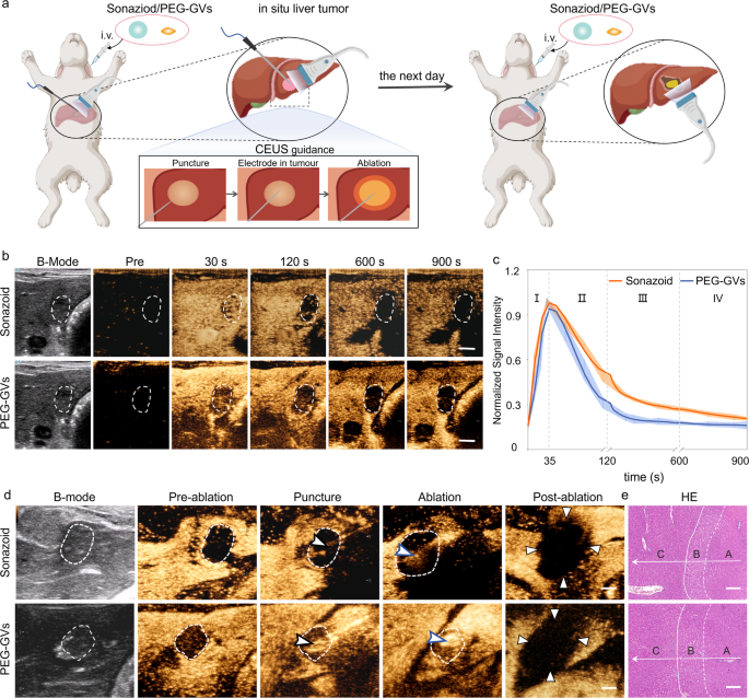
Given the good potential of PEG-GVs in ultrasound imaging of the liver and liver tumors, we additional investigated their position in guiding radiofrequency ablation (RFA) in an in situ rabbit liver tumor mannequin (Determine S8a). As is well-known, the distinct show of tumor boundaries is important for figuring out tumor ablation extent. Nevertheless, the tumor boundaries normally can’t be clearly recognized on grayscale ultrasound. In distinction, CEUS can exactly find the lesion, information the puncture, and promptly consider the therapeutic impact throughout ablation. Contemplating that Sonazoid has been extensively utilized in guiding RFA ablation, we carried out a comparability between Sonazoid and PEG-GVs for guiding ablation of liver tumor [34, 35]. Sonazoid or PEG-GVs had been administered intravenously to rabbits with orthotopic VX2 tumors after which carried out the ultrasound-guided RFA, adopted by a second administration subsequent day (Fig. 6a). As illustrated in Fig. 6b, each PEG-GVs and Sonazoid exhibited a broader spectrum of tumors in comparison with their two-dimensional B-mode pictures. Notably, the tumor vary displayed by PEG-GVs was even bigger and remained sturdy over time. The normalized sign depth curves indicated that each PEG-GVs and Sonazoid exhibited fast perfusion and regression in early liver tumors (Fig. 6c). Nevertheless, PEG-GVs retained within the surrounding liver tissue for an extended interval, producing extra distinct tumor boundaries. The tumor boundaries displayed by PEG-GVs make it extra acceptable for guiding exact tumor ablation, particularly for guiding punctures and observing the ablation vary by ultrasound in a real-time method, as depicted in Fig. 6d. HE staining clearly revealed regular liver tissue, peritumoral edematous liver tissue, and coagulated necrotic liver tumor tissue after RFA, indicating that full ablation was achieved in each teams (Fig. 6e and Determine S8b). These outcomes counsel that PEG-GVs performs an analogous position to Sonazoid in guiding RFA of liver tumors.
Position of PEG-GVs in guiding RFA of liver tumor. a, Diagram of and CEUS-guided RFA therapy after administration of Sonazoid or PEG-GVs in rabbits in situ liver tumor. b–c, CEUS pictures in numerous phases of rabbits in situ liver tumor (b) and normalized sign depth curves (c), (n = 5). Scale bar: 5 mm. d, B-mode ultrasound pictures of preoperative tumor, revealing the unclear tumor boundary (white dotted circles). The tumor boundary might be seen in CEUS picture until the portal venous part (white dotted circles). With the CEUS steering, monitoring puncture, radiofrequency electrodes (black arrowheads) had been simply inserted into the index tumor. The electrode needle was turned on and ablation had been carried out (blue arrowheads). The CEUS was repeated after RFA ablation. Triangle factors to the world of non-enhancement within the early arterial part, indicating a necrotic tumor instantly after RFA. (n = 3). Scale bar: 5 mm. e, HE staining of the ablated tissue. A, B, C stand for coagulated necrotic liver tumor after RFA, peritumoral edematous liver tissue and regular liver tissue, respectively (n = 3). Scale bar: 200 μm.
CEUS imaging and biosafety of PEG-GVs in macaque
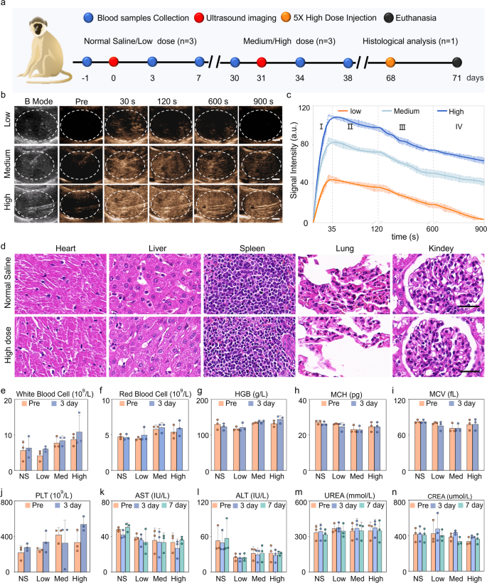
Macaques are extremely just like people by way of tissue construction, physiology, and metabolism. Therefore, we additional investigated the imaging efficiency of PEG-GVs in macaques. The experimental course of is depicted in Fig. 7a. Determine 7b and c demonstrated that every dose of PEG-GVs might yield a fast enhancement, reaching a peak about 35 s after systemic administration, then step by step decreased over time. With the rise of bubble doses, the distinction sign intensities elevated and persevered for an extended length. Notably, 15-min length was exhibited for medium and excessive dose teams. Quantitative knowledge indicated that the distinction sign intensities of three group at peak achieved 48.44 ± 3.70 a.u, 86.15 ± 3.11 a.u, 113.8 ± 3.66 a.u., respectively. It’s value noting that medium and excessive dose teams remained almost half of the sign intensities after 900 s following the injection of PEG-GVs (Fig. 7c). To confirm the in vivo biosafety of PEG-GVs, histological evaluation of main organs and serum biochemical assays had been carried out on the endpoint. HE staining knowledge indicated that there was no important harm to the guts, lung, kidney, and liver within the excessive dose macaque group (Fig. 7d), just like the outcomes obtained in mice (Determine S9). Assessments of blood samples, together with liver perform, kidney perform, and blood rely, additional indicated that PEG-GVs didn’t trigger important uncomfortable side effects (Fig. 7e-n, Determine S10). Subsequently, PEG-GVs, as a novel ultrasound distinction agent, would possibly maintain broad prospects for future scientific purposes.
Liver CEUS imaging and biosafety of PEG-GVs in macaque. a, Schematic diagram of biosafety evaluation in macaques. b-c, CEUS pictures of macaque liver (b) and the sign depth curves of the three ultrasonic brokers in 4 phases (c). d, HE staining of main organs of macaques acquired with regular saline or high-dose PEG-GVs. e-n. Hematological detection of every index earlier than and after systemic administration of regular saline or PEG-GVs.

