Synthesis and characterization of HA-CUR NPs
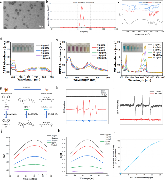
HA-CUR NPs had been synthesized through esterification between the carboxyl group of HA and the hydroxy group of CUR via a simple and reliable technique. Particularly, CUR was dissolved in a response system containing HA, DCC, and DMAP to type HA-CUR NPs. After 24 h of stirring, the ensuing combination turned darkish, suggesting the efficient mixture of HA and CUR. The combination was subsequently dialyzed and centrifuged to acquire HA-CUR NPs with superior aqueous dispersion. The micromorphology of the HA-CUR NPs was noticed through TEM. As proven in Fig. 1a, HA-CUR NPs had been usually within the type of uniformly distributed ultrafine particles with a dimension of twenty-two.4 ± 8.7 nm. The common hydrodynamic dimension of the HA-CUR NPs was subsequently measured through DLS and was similar to the end result decided through TEM (Fig. 1b). We subsequently decided the construction of the HA-CUR NPs by measuring their 1H NMR spectra. As proven in Determine S1, the peaks of HA-CUR at 1.5–2.0 ppm and 6.5–7.0 ppm had been attributed to the acetyl peak of HA (- NHCOCH3) and the fragrant proton of CUR, respectively. Moreover, the infrared intensities of the HA, CUR, and HA-CUR NPs had been analyzed through FTIR to confirm that the HA-CUR NPs had been efficiently synthesized. As proven in Fig. 1c, in contrast with the infrared spectra of HA and CUR, the obvious absorption peak at 3327 cm-1 was the absorption band of the -OH group within the curcumin construction; the plain absorption peak at 2930 cm-1 was the absorption band of the double C = O group in curcumin; the brand new absorption peak at 1576 cm-1 was attributed to the vibration of the C = C throughout the benzene ring of curcumin; and the brand new absorption peak at 1732 cm-1 was the absorption band of the newly fashioned C = O double bond within the ester. Furthermore, the HA-CUR NPs had been dissolved in three solvents: water, ethanol and 0.9% NaCl. The UV‒vis absorbance spectra indicated that there was no marked change at 0 and seven days (Determine S2). These outcomes had been in line with the findings of Manju S, indicating the profitable synthesis of HA-CUR NPs.
Characterization, ROS scavenging skill, and enzyme-mimicking actions of HA-CUR NPs. (a) Transmission electron microscopy photographs of HA-CUR NPs. (b) Dynamic mild scattering information of HA-CUR NPs. (c) FTIR spectra of the HA, CUR, and HA-CUR NP samples. (d) UV‒Vis absorbance exhibiting the ABTS radical scavenging skill of HA-CUR NPs. (e) UV‒Vis absorbance exhibiting the DPPH radical scavenging skill of HA-CUR NPs. (f) UV‒Vis absorbance picture illustrating the flexibility of HA-CUR NPs to scavenge ·OH to guard MB. (g) Illustration of the ROS scavenging course of. (h) ESR spectra of DMPO indicating ·OH seize with or with out HA-CUR NPs. (i) ESR spectra of TEMPO indicating single oxygen seize with or with out HA-CUR NPs. (j) SOD-like exercise of HA-CUR NPs. (WST-8 equipment assay) (n = 3 for every group). (ok) GSH-like exercise of HA-CUR NPs. (n = 3 for every group). (l) CAT-like exercise of HA-CUR NPs (n = 3 for every group)
Radical scavenging and antioxidant capability of HA-CUR NPs
CUR has robust antioxidant properties, however its low aqueous solubility significantly limits its widespread software. Right here, we explored the free radical scavenging skill and antioxidant capability of HA-CUR NPs. First, the ABTS and DPPH radical assays, that are two radical fashions broadly used for quantifying the antioxidant capability of organic samples, had been adopted to check the novel scavenging skill [18, 19]. The capability of HA-CUR NPs to scavenge ABTS and DPPH radicals was evaluated by measuring the absorbances at 734 and 517 nm, respectively. Because the focus of HA-CUR NPs elevated, the colour step by step decreased, and decreased absorbance values had been noticed (Fig. 1d and e), suggesting the robust ABTS and DPPH radical scavenging skills of the HA-CUR NPs. Moreover, the OH radical (·OH) is a physiologically associated radical and is concerned in varied sorts of mobile and histopathological harm. The power of HA-CUR NPs to scavenge ·OH was assessed through the methylene blue (MB) assay. Our findings revealed that with the safety of HA-CUR NPs, MB remained blue or pale barely, indicating that HA-CUR NPs had a robust scavenging skill in opposition to ·OH (Fig. 1f). The precise mechanisms of those three assays are schematically introduced in Fig. 1g. As well as, 5,5-dimethyl-1-pyrroline N-oxide (DMPO) and a pair of,2,6,6-tetramethylpiperidine-1-oxyl (TEMPO) had been employed to lure •OH and single oxygen, respectively. The authors evaluated the capability of HA-CUR NPs to scavenge free radicals produced by the Fenton response via electron spin resonance (ESR) spectroscopy. A decreased ESR amplitude was noticed after remedy with the HA-CUR NPs, suggesting the environment friendly scavenging skill of the HA-CUR NPs for ·OH and single oxygen (Fig. 1h and that i). In abstract, these findings point out that HA-CUR NPs could also be efficient ROS scavengers due to their robust antioxidant properties.
Mimetic actions of three antioxidant enzymes within the HA-CUR NPs
The results of HA-CUR NPs on SOD, GSH, and CAT exercise had been additional explored. SOD, an antioxidant enzyme that decomposes superoxide radicals into hydrogen peroxide and oxygen, has been demonstrated to exert outstanding therapeutic results in uveitis [20]. Moreover, GSH is a broadly current tripeptide thiol and one other essential intracellular and extracellular antioxidant that performs many essential roles in mobile sign transduction processes [21]. H2O2 will be eradicated from the physique by decomposition into H2O and O2 by CAT, an iron porphyrin-binding enzyme that performs a significant protecting position in organisms. As proven in Fig. 1j, ok, and l, the HA-CUR NPs demonstrated SOD, GSH, and CAT mimetic results in a dose-dependent method, suggesting that the HA-CUR NPs have glorious antioxidant results.
Mobile uptake habits of HA-CUR NPs and in vitro cytotoxicity of HA-CUR NPs
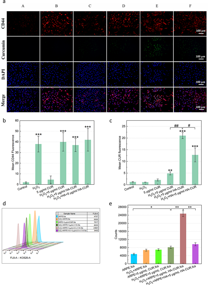
The pathogenesis and development of uveitis are carefully related to the impairment of ocular immune privilege, which can be attributable to destruction of the integrity of the RPE [22]. Flourescence microscopy was employed to guage the flexibility of HA-CUR NPs to focus on ARPE-19 cells by figuring out their internalization of HA-CUR NPs and free CUR pretreatment with or with out H2O2. CUR is represented by the inexperienced fluorescence sign, whereas the CD44 receptor is represented by the purple sign. ARPE-19 cells that weren’t handled with H2O2 assimilated minimal quantities of free CUR, as demonstrated in Fig. 2a, and the quantitative evaluation outcomes are proven in Fig. 2b and c. The mobile uptake habits of HA-CUR NPs was extra simply noticed in ARPE cells pretreated with H2O2 than in these pretreated with free CUR. The next observations had been made to establish whether or not the elevated mobile absorption of HA-CUR NPs by ARPE-19 cells pretreated with H2O2 was a results of their skill to focus on the CD44 receptor. Initially, pretreatment of ARPE-19 cells with H2O2 led to better expression of the CD44 receptor than that in untreated ARPE-19 cells. These findings point out that the expression of the CD44 receptor could also be promoted by extreme oxidative stress. ARPE-19 cells had been then handled with HA or HA-CUR NPs following H2O2 pretreatment. The absorption of HA-CUR NPs by cells clearly decreased, probably on account of the aggressive binding of HA to the CD44 receptor, which inhibited NP-CD44 receptor binding. The outcomes had been additional verified via movement cytometry (Fig. 2d). MTT assays revealed that the exercise of ARPE-19 cells was diminished by incubation with greater concentrations of HA-CUR NPs (Determine S3a-b). The relative survival charge of the cells and the proportion of these with a standard morphology decreased (Determine S3c). In abstract, these information point out that the CD44 receptor concentrating on of HA-CUR NPs might promote the mobile uptake of HA-CUR NPs resulting from CD44 receptor overexpression in ARPE-19 cells attributable to extreme oxidative stress.
(a) Fluorescence photographs exhibiting CD44 and CUR expression in several teams. A: Management; B: 500 µmol/L H2O2-treated APRE-19 cells for 40 min; C: 5 µg/mL CUR-treated APRE-19 cells for 12 h; D: 500 µmol/L H2O2-treated APRE-19 cells for 40 min + 5 µg/mL CUR-treated APRE-19 cells for 12 h; E: 500 µmol/L H2O2-treated APRE-19 cells for 40 min + 5 µg/mL HA-CUR NP-treated APRE-19 cells for 12 h; F: 500 µmol/L H2O2-treated APRE-19 cells for 40 min + 5 µg/mL HA-treated APRE-19 cells for 4 h + 5 µg/mL HA-CUR NP-treated APRE-19 cells for 12 h. (b) Quantification of the fluorescence depth of CD44 in several teams. (c) Quantification of the fluorescence depth of CUR in several teams. (d, e) Stream cytometry evaluation of the fluorescence depth of CUR in several teams. Observe: */#, P < 0.05; **/##, P < 0.01; ***/###, P < 0.001
HA-CUR NPs attenuated oxidative stress and inflammatory responses in ARPE-19 cells
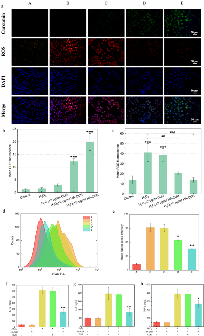
Inspired by their glorious mobile uptake, HA-CUR NPs had been evaluated for his or her skill to safeguard ARPE-19 cells from oxidative stress attributable to H2O2. DCFH-DA staining was carried out to visualise intracellular ROS after completely different interventions. The inexperienced fluorescence sign signifies CUR, and the purple fluorescence sign represents ROS. In contrast with people who weren’t handled with H2O2, ARPE-19 cells that had been pretreated with H2O2 introduced a purple fluorescence sign, which steered that the intracellular ROS ranges had been elevated, as demonstrated by Fig. 3a and the outcomes of the quantitative evaluation proven in Fig. 3b and c. When the cells had been cocultured with free CUR, vital intracellular ROS and very low mobile uptake of CUR had been detected. Nonetheless, the purple fluorescence sign decreased considerably, and the inexperienced fluorescence sign elevated when H2O2-pretreated ARPE-19 cells had been cocultured with HA-CUR NPs, suggesting that HA-CUR NPs may cause a dose-dependent discount in ROS.
(a) Fluorescence photographs of ARPE-19 cells stained with DFCH-DA and 4′,6-diamidino-2-phenylindole (DAPI) after completely different therapies. A: Management; B: 500 µmol/L H2O2-treated APRE-19 cells for 40 min; C: 500 µmol/L H2O2-treated APRE-19 cells for 40 min + 2 µg/L CUR-treated APRE-19 cells for 12 h; D: 500 µmol/L H2O2-treated APRE-19 cells for 40 min + 2 µg/mL HA-CUR NP-treated APRE-19 cells for 12 h; E: 500 µmol/L H2O2-treated APRE-19 cells for 40 min + 5 µg/mL HA-CUR NP-treated APRE-19 cells for 12 h. (b) Quantification of the fluorescence depth of CUR in several teams. (c) Quantification of the fluorescence depth of ROS in several teams. (d) Stream cytometry quantification of ARPE-19 cells stained with DFCH-DA after completely different therapies. (e) Imply fluorescence depth calculated from the movement cytometry information in d; (f) expression degree of IL-1β in several teams; (g) expression degree of IL-6 in several teams; (h) expression degree of TNF-α in several teams. Observe: */#, P < 0.05; **/##, P < 0.01; ***/###, P < 0.001
These findings had been additional verified through movement cytometry (Fig. 3d and e). Moreover, H2O2-pretreated ARPE-19 cells exhibited markedly elevated manufacturing of inflammatory cytokines, together with IL-1β, IL-6 and TNF-α, which had been downregulated by HA-CUR NPs however not by CUR (Fig. 3f, g and h). These outcomes collectively point out that HA-CUR NPs might mitigate ARPE-19 cell oxidative stress and the inflammatory response.
HA-CUR NPs attenuated pathological development, relieved microvascular harm, and controlled fundus blood movement in vivo
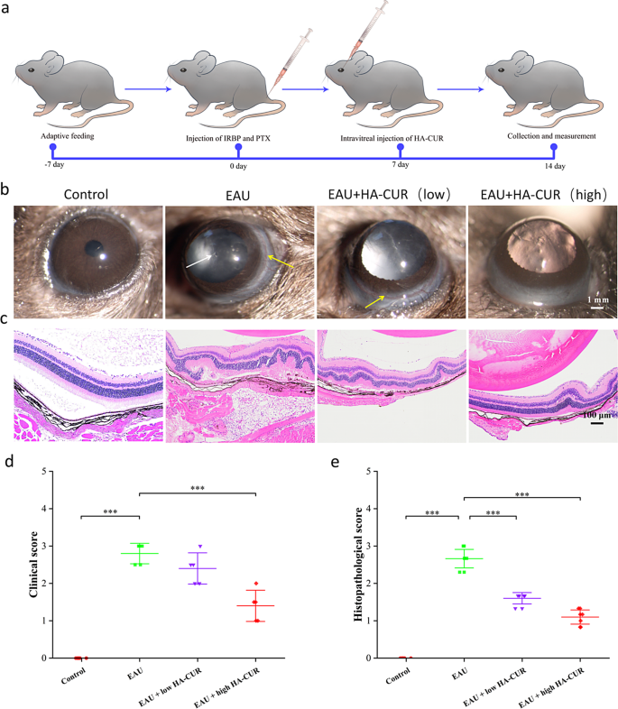
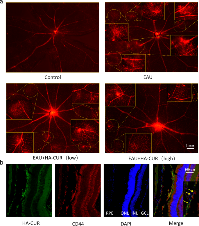
EAU mice’s medical signs and retinal tissue pathological adjustments had been graded by severity in line with earlier requirements [23, 24]. On the idea of the wonderful mobile uptake habits and skill of HA-CUR NPs to guard in opposition to oxidative stress harm in vitro, we researched the therapeutic efficacy of HA-CUR NPs in vivo (Fig. 4a). In contrast with regular management rats, EAU mice introduced vital irritation within the anterior chamber and extreme ciliary and conjunctival hyperemia, whereas EAU mice handled with an intravitreal injection of excessive HA-CUR NPs introduced relieved signs (Fig. 4b). H&E staining revealed typical histopathological indicators within the EAU mannequin, together with considerably elevated retinal folds and elevated inflammatory cells, which had been markedly attenuated within the EAU mannequin mice handled with the intravitreal injection of excessive concentrations of HA-CUR NPs (Fig. 4c). Medical and histopathological scores had been additionally assessed (Fig. 4d and e). The results of HA-CUR NPs on the regulation of retinal vascularity and blood movement in vivo had been additionally totally investigated. Evans blue serves as a diazo dye that shortly and irreversibly binds to plasma ALB and subsequently can be utilized as a extremely environment friendly retinal angiographic agent. As proven within the retinal vascular community photographs obtained from Evans blue staining in Fig. 5a, huge vascular leakage websites had been noticed within the retinas of the EAU mice, and this leakage was markedly alleviated within the EAU mice handled with the intravitreal injection of excessive concentrations of HA-CUR NPs (Determine S4). Conversely, signs weren’t considerably ameliorated in EAU mannequin mice following the intravitreal injection of low concentrations of HA-CUR NPs. Moreover, immunohistochemistry was applied to guage the concentrating on of RPE cells by HA-CUR NPs. As proven in Fig. 5b, HA-CUR NPs might be noticed within the cytoplasm of RPE cells. The outcomes revealed the colocalization of HA-CUR NPs and CD44-positive RPE cells.
(a) Illustration of the experimental process used to determine the experimental autoimmune uveitis (EAU) mannequin. (b) Medical manifestations of retinas within the completely different teams had been noticed through a slit lamp. The white arrows point out anterior chamber irritation; the yellow arrows point out conjunctival and ciliary congestion. (c) HE-stained histopathological photographs of retinas from the completely different teams. (d) Medical rating based mostly on the leads to (b). (e) Histopathological rating based mostly on the leads to (C). Observe: *, P < 0.05; **, P < 0.01; ***, P < 0.001
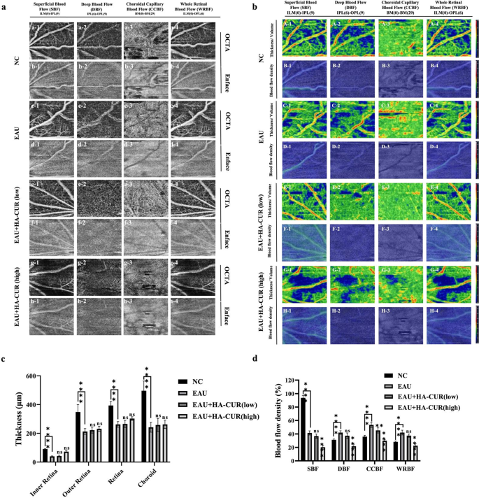
OCTA, a noninvasive fundus vascular imaging expertise that’s progressive, has been extensively employed to diagnose fundus issues, together with diabetic retinopathy and uveitis. In contrast to conventional strategies, OCTA doesn’t require the intravenous software of dye, stopping fluorescein leakage [25]. We employed OCTA imaging to quantify the thickness and density of blood movement in every fundus layer in distinct teams of mice. Determine 6a reveals the superficial blood movement (SBF), deep blood movement (DBF), choroidal capillary blood movement (CCBF), and entire retinal blood movement (WRBF) of the fundus of the varied teams. In distinction to NC mice, EAU mice introduced typical retinal vascular morphological anomalies. Conversely, the retinal vascular networks of EAU mannequin mice had been regular following intravitreal injections of each high and low concentrations of HA-CUR NPs. Determine 6b reveals the OCTA fundus blood movement and thickness/quantity maps. In contrast with NC mice, EAU mice introduced extra impaired blood movement alerts and uneven vessel morphology throughout the entire retinal vascular community, indicating vital vascular dysfunction within the fundus of the EAU mice. In distinction, these irregular signs had been alleviated in EAU mannequin mice by intravitreal injections of each high and low concentrations of HA-CUR NPs. The quantitative evaluation of the SBF, DBF, CCBF, and WRBF layers of the fundus, as illustrated in Fig. 6c and d, revealed a marked lower in thickness within the EAU group in contrast with the NC group. Each the EAU mice handled with the intravitreal injection of low- and high-HA-CUR NPs subsequently exhibited outstanding enhancements to comparatively regular ranges. These outcomes point out that HA-CUR NP remedy markedly attenuated pathological development, relieved microvascular leakage, and controlled fundus blood movement; subsequently, HA-CUR NPs function efficient brokers for retinal safety in EAU.
Optical coherence tomography angiography (OCTA) imaging, blood movement, and thickness of a number of layers of OCTA within the fundus. (a) Superficial blood movement (SBF) (a1-h1), deep blood movement (DBF) (a2-h2), choroidal capillary blood movement (CCBF) (a3-h3), and entire retinal blood movement (WRBF) (a4-h4) of the OCTA panorama in several teams. (b) OCTA fundus thickness/quantity (A, C, E, G) and blood movement (B, D, F, H) maps in several teams. (c) Quantification of the thickness of the SBF, DBF, CCBF, and WRBF retinal layers in several teams. (d) Quantification of blood movement within the SBF, DBF, CCBF, and WRBF retinal layers in several teams. Asterisks point out a major distinction. Observe: *, P < 0.05; **, P < 0.01; ***, P < 0.001; ****, P < 0.0001
HA-CUR NPs mitigated oxidative stress and diminished inflammatory responses
HA-CUR NPs have been proven to mitigate oxidative stress in vitro; thus, we investigated whether or not HA-CUR NPs exert related results in vivo. MDA is normally thought to be a marker for lipid peroxidation and oxidative stress [26], whereas SOD is a metalloenzyme that safeguards the organism in opposition to oxidative stress [27]. The redox state of uveitis is usually recommended to be imbalanced, as evidenced by the considerably greater intraocular MDA and diminished intraocular SOD in EAU animals than in regular controls (Fig. 7a-j). Happily, the administration of HA-CUR NPs dose-dependently decreased excessive MDA ranges and elevated low SOD ranges in EAU mice, whereas remedy with free CUR had a minimal impact on these indicators. Moreover, on condition that IL-17, IFN-γ and TNF-α are attribute inflammatory cytokines that take part within the pathogenesis and growth of uveitis [28], we additional examined the expression of those three cytokines within the completely different teams. In contrast with NC mice, EAU mice introduced markedly better expression ranges of intraocular IL-17, IFN-γ and TNF-α, which had been decreased in a dose-dependent method by remedy with HA-CUR NPs fairly than free CUR. Moreover, we quantified the expression of the CD44 receptor in response to the extreme oxidative stress that was induced in vitro (Fig. 7ok, l). The CD44 receptor expression of the EAU mice exceeded that of the traditional controls, suggesting a probably extra intense focused interplay between HA and the receptor. The mixture of CUR and HA might enhance CUR bioavailability by concentrating on HA to the CD44 receptor, decreasing oxidative stress and inflammatory responses, on condition that HA-CUR NPs are extra efficacious than free CUR is. Contemplating that HA-CUR NPs are considerably extra efficacious than free CUR is, one cheap conclusion will be drawn that the mix of CUR and HA may markedly enhance the bioavailability of CUR, which can be mediated by the particular concentrating on of HA to the CD44 receptor, thereby effectively attenuating extreme inflammatory responses and oxidative stress.
Quantification of the serum MDA (a), SOD (b), TNF-α (c), IFN-γ (d), and IL-17 (e) ranges and the intraocular MDA (f), SOD (g), TNF-α (h), IFN-γ (i), and IL-17 (j) ranges in several teams. (ok, l) Expression of the CD44 receptor in management and EAU rats. Observe: */#, P < 0.05; **/##, P < 0.01; ***/###, P < 0.001
HA-CUR NPs exerted anti-inflammatory results by activating the Keap1/Nrf2/HO-1 signaling pathway
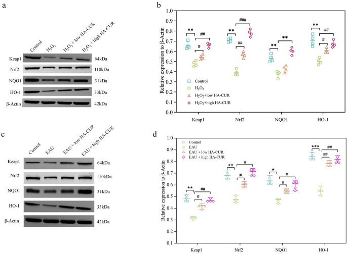
By figuring out the potential mechanism behind the outstanding oxidative stress safety of HA-CUR NPs, we might be able to higher perceive the therapeutic advantages of HA-CUR NPs and optimize their medical software. To additional discover the molecular mechanism of HA-CUR NPs, we carried out transcriptome sequencing evaluation on ARPE-19 cells handled with completely different interventions. The Molecular Signatures Database C2 database was used to carry out GSEA on the DEGs, and the highest 20 outcomes are illustrated in Fig. 8a. Nearly all of genes whose expression was considerably upregulated by HA-CUR NPs had been related to irritation, immunology, and oxidative stress, together with REACTOME_KEAP1_NFE2L2_PATHWAY, a traditional pathway that’s concerned within the antioxidative stress response and safeguards cells from oxidative harm. Extra enrichment evaluation confirmed the activation of the Keap1/Nrf2 signaling pathway (Fig. 8b and c). A gene expression heatmap of the Keap1/Nrf2 signaling pathway revealed that a number of genes within the Keap1/Nrf2 signaling pathway, together with GSTM5, GSTO2, MGST1, KEAP1, HMOX1, and NFE2L2, had been upregulated by the HA-CUR NPs (Fig. 8d and e), which promoted the expression of anti-inflammatory and antioxidant-related proteins to withstand oxidative stress harm. As well as, the highest 5 potential signaling pathways and the highest 6 DEGs revealed by transcriptome sequencing evaluation are proven in Fig. 8f. As one of the very important antioxidant axes in physiological and pathological processes, the activation of Nrf2 signaling may contribute to a collection of antioxidant responses, thereby defending cells in opposition to oxidative stress harm [29, 30]. Particularly, Nrf2 is a transcription component that promotes the transcription and expression of varied main antioxidative enzymes, together with heme oxygenase-1 (OH-1) and NAD(P)H quinone oxidoreductase 1 (NQO1), by binding to antioxidation stress response websites [30, 31]. In quite a few cell strains, together with RPE cells, pure merchandise have been proven to change the Keap1/Nrf2/HO-1 signaling pathway [32]. Notably, Nrf2 was discovered to be carefully associated to the event of uveitis, serving as a possible intervention goal within the modulation of redox hemostasis [33]. Western blotting was used to find out whether or not HA-induced CUR NPs can safeguard ARPE-19 cells and EAU mannequin mice from oxidative stress-induced harm by activating the Keap1/Nrf2/HO-1 signaling pathway. The protein expression ranges of Keap1, Nrf2, HO-1, and NQO1 in ARPE-19 cells pretreated with hydrogen peroxide had been considerably decrease than these within the management group. Nonetheless, the H2O2-induced results on the Keap1/Nrf2/HO-1 signaling pathway had been reversed by HA-CUR NPs in a dose-dependent method, as illustrated in Fig. 9a and b. Moreover, in distinction to these within the management group, the protein expression ranges of Keap1, Nrf2, HO-1, and NQO1 within the EAU group had been considerably decrease. Then again, HACUR NPs had been nonetheless able to reversing the adjustments in protein expression ranges in a dose-dependent method (Fig. 9c and d). These findings point out that Keap1/Nrf2/HO-1 induces the protecting affect of the HA CUR NPs. These outcomes counsel that the Keap1/Nrf2/HO-1 signaling pathway contributes considerably to the anti-inflammatory impact of the HA CUR NPs. Curcumin is a pure activator of Nrf2 [34]. Mechanistic research have proven that curcumin induces conformational adjustments in Keap1 and the ubiquitination of Nrf2 by binding to Keap1Cys151 [35]. Nonetheless, the consequences of curcumin on the protein expression of Nrf2 in cells subjected to oxidative stress should not solely constant. Liu et al. confirmed that pretreatment with curcumin didn’t enhance the expression of complete Nrf2 in cells subjected to oxidative stress stimulation [36]. These findings are inconsistent with our findings. Nonetheless, some research have proven that curcumin can enhance the protein expression of Nrf2 in cells subjected to numerous oxidative stresses [35, 37]. The focus of curcumin used and the length of its motion could also be key elements influencing the extent of Nrf2 expression.
(a) The highest 20 outcomes of GSEA enrichment evaluation of the DEGs through the Molecular Signatures Database C2 database. (b, c) Working enrichment rating of the Keap1/Nrf2 signaling pathway. (d, e) Gene expression heatmap of the Keap1/Nrf2 signaling pathway regulated by HA-CUR NPs. (f) Protein ranges of Keap1, Nrf2, HO-1, and NQO1 in ARPE-19 cells after completely different therapies. h, i) Protein ranges of Keap1, Nrf2, HO‐1, and NQO1 in rats in several teams
Evaluation of biocompatibility in vivo
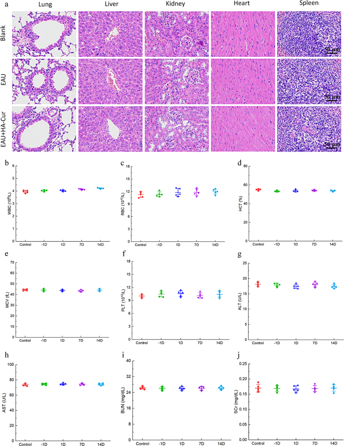
The biocompatibility of HA-CUR NPs in vivo was assessed via H&E staining of essential organs, together with the center, liver, spleen, lung, and kidney. As proven in Fig. 10a-j, no outstanding lesions, hemorrhage, or necrosis had been detected in these main organs. No substantial adjustments in liver or renal operate had been noticed in any of the mice within the current examine, as decided by ALT, AST, BUN, and SCR within the serum. The WBC, RBC, PLT, MCV, and HCT had been additionally measured and demonstrated no vital adjustments. No apparent lesions had been discovered within the retinal tissue after the HA-CUR NPs had been injected into the attention. (Determine S5a). We additional examined three inflammatory cytokines to guage the toxicity of HA-CUR NPs to intraocular tissues. As proven in Determine S5b, the expression ranges of IL-17, IFN-γ and TNF-α within the eyes of the mice injected with HA-CUR NPs didn’t considerably differ from these within the management group. Due to this fact, the HA-CUR NPs synthesized within the current examine have the potential to function protected and efficacious brokers for stopping the development of EAU.
In vivo biocompatibility of HA-CUR NPs. (a) Pictures of H&E-stained coronary heart, liver, spleen, lung, and kidney tissues from the completely different teams. (b–f) Quantification of white blood cells (WBCs), purple blood cells (RBCs), platelets (PLTs), imply corpuscular quantity (MCV), and hematocrit (HCT) within the completely different teams. (g–j) Serum alanine aminotransferase (ALT), aspartate aminotransferase (AST), blood urea nitrogen (BUN), and serum creatinine (SCR) ranges within the completely different teams

