Complete characterization of LMWH-LA conjugate and its self-assembled nanoparticles
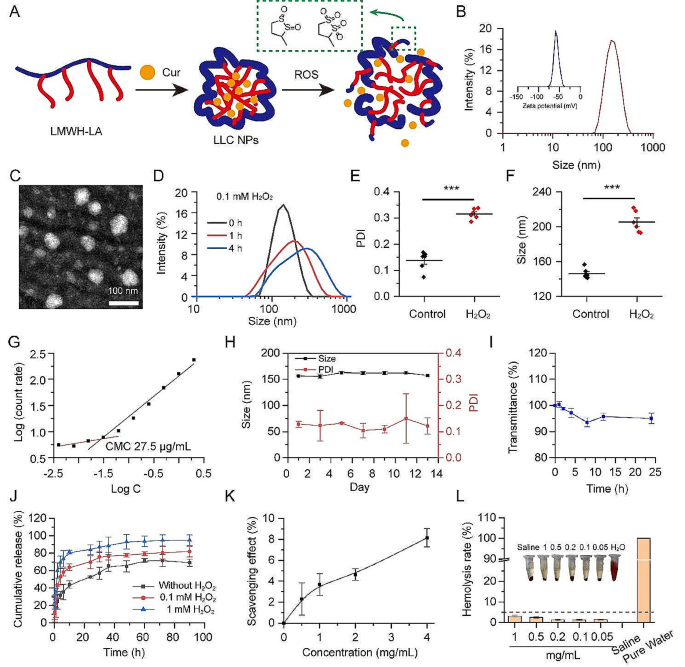
Inflammatory cells are concerned in lots of key hyperlinks within the improvement of atherosclerosis, resembling adhesion of monocytes, polarization of macrophages, and secretion of ROS and inflammatory elements. In our earlier research, nanoparticles with LMWH as shells may competitively bind P-selectin on injured vascular endothelial cells with monocytes, thereby lowering monocytes’ adherence to vascular endothelium [33]. The excessive ROS degree on plaque additionally guided us to rationally select LA as a ROS set off and scavenger. With a purpose to obtain cascade inhibition of plaque irritation, LA was conjugated to the LMWH chain to type a multifunctional amphipathic drug service for atherosclerosis focused remedy. The hydrophobic LA section can reply to the excessive ROS within the plaque and rework into hydrophilic, thereby releasing the encapsulated medicine (Fig. 2A). The chemical buildings obtained had been analyzed utilizing proton nuclear magnetic resonance ([1]H NMR) spectra and Fourier rework infrared (FT-IR) spectroscopy (Fig. S1–2). By way of thermogravimetric evaluation (TGA), it was decided that the content material of lipoic acid (LA) throughout the LMWH-LA conjugate was round 20% (w/w) (Fig. S3).
Due to their amphiphilic chemical buildings, LMWH-LA is able to self-assembling into micelles, generally known as LL NPs, that includes the LMWH chain because the hydrophilic outer shell and the LA unit because the hydrophobic inside core. Cur might be encapsulated inside these micelles, forming LLC NPs by way of hydrophobic interactions. Dynamic gentle scattering (DLS) measurements revealed that the LL NPs have a median dimension of about 143 nm (Fig. S4). Upon incorporating Cur, the particle dimension of the LLC NPs expanded to 160 nm (Fig. 2B). The LLC NPs exhibited a unfavourable ζ potential of -50 mV. The loading capability for Cur and the encapsulation effectivity throughout the nanoparticles had been measured to be 7.8% and 85%, respectively. TEM confirmed that LLC NPs had been spherical in morphology (Fig. 2C). To substantiate the ROS sensitivity, the dimensions distribution and polydispersity index (PDI) of LL NPs incubated with 0.1 mM H2O2 had been evaluated. The dimensions considerably elevated and the dimensions distribution considerably widened after 4 h stimulation, displaying an apparent ROS sensitivity of LL NPs (Fig. 2D, E, F). The great stability of NPs was a prerequisite for coming into medical analysis. Firstly, the CMC of the LL NPs was evaluated by dynamic gentle scattering. In Fig. 2G, the CMC was calculated as 27.5 µg/mL. Subsequent, the storage stability of LLC NPs at 4 °C was assessed by monitoring adjustments in nanoparticle dimension and PDI, with no notable alterations in both parameter noticed over a 13-day interval, demonstrating their favorable storage stability (Fig. 2H). Then, the soundness of NPs in serum was evaluated by incubating them with 50% fetal bovine serum (FBS) at 37 °C. Over a 24-hour interval, there was no vital variation in transmission, indicating that the LLC NPs maintained their stability in 50% FBS (Fig. 2I). Moreover, the discharge sample of the drug from LLC NPs was analyzed in PBS (containing 0, 0.1 and 1 mM H2O2) by inspecting the Curcumin launch profiles. The presence of H2O2 would speed up the discharge of Cur, and the upper H2O2 focus, the sooner the drug launch, which was associated to the change in hydrophobicity of the micelle core (Fig. 2J). Earlier research have identified LA was thought to be potent mobile oxidation regulators with extraordinary antioxidant properties [34]. So, it’s essential to discover the antioxidant properties of LA grafted onto LMWH. As proven in Fig. 2Okay, LL NPs revealed good antioxidant properties, and its antioxidant capability was positively correlated with the focus. Furthermore, preliminary outcomes from hemolysis assessments indicated that the hemolysis charges of LL NPs didn’t exceed 5% at a focus of 1000 µg/mL and the pink blood cells confirmed regular morphology, indicating that LL NPs posed no hemolysis threat. (Fig. 2L, Fig. S5).
Total, the elements of LMWH-LA had been all derived from medical medicine, and its construction was clear. The NPs shaped by LMWH-LA have good stability, ROS sensitivity, antioxidant capability and good hemocompatibility, which supplied a foundation for subsequent anti-atherosclerosis remedy.
The LMWH-LA conjugates self-assembles right into a secure, ROS delicate micelles with controllable launch and good blood compatibility. (A) Schematic diagram of self-assembly and ROS response of LLC NPs. (B) Particle dimension and zeta potential of LLC NPs. (C) TEM photographs of LLC NPs (scale bar = 100 nm). (D) Particle dimension distribution of LL NPs beneath ROS stimulation for 1 h and 4 h. (E, F) The adjustments in PDI and particle dimension beneath ROS stimulation for 4 h. means ± SD, n = 6. (G) The CMC of LL NPs. (H) The steadiness of LLC NPs over a 15-day interval at 4 °C. Means ± SD, n = 3. (I) The steadiness of NPs in serum for twenty-four h when incubated with 50% FBS at 37 °C. Means ± SD, n = 3. (J) Cumulative Cur launch of LLC NPs in PBS pH 7.4 beneath totally different ROS concentrations. Means ± SD, n = 3. (Okay) The ROS scavenging impact of LL NPs. means ± SD, n = 3. (L) The ratio of hemolysis in pink blood cells uncovered to LL NPs at totally different concentrations. Means ± SD, n = 3. Embedded are photographs of pink blood cell suspension
Suppression of the vascular irritation cascade by way of in vitro NPs intervention
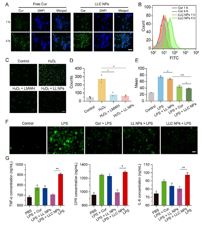
Macrophages play an necessary position in plaque irritation. On this half, the cytotoxicity and mobile uptake of NPs in RAW264.7 cells had been investigated by way of in vitro experiments. LL NPs had no toxicity to RAW264.7 cells in Fig. S6. Determine 3A depicted the mobile uptake. Following a 1-hour incubation interval with LLC NPs, inexperienced fluorescence was noticed surrounding the nucleus of RAW264.7 cells, demonstrating the speedy entry of LLC NPs into RAW264.7 cells. When the co-incubation time reached 4 h, stronger inexperienced fluorescence was noticed, indicating a time-dependent uptake of NPs by RAW264.7 cells. In distinction, free Cur confirmed a decrease uptake effectivity by RAW264.7 cells as a consequence of its poor water solubility. As well as, circulate cytometry additionally revealed that LLC NPs had been extra simply taken up by cells than free Cur (Fig. 3B).
Monocyte adhesion to compromised endothelial cells inside blood vessels marks the preliminary section of plaque-related irritation. Herein, HUVECs had been handled with H2O2 to create an inflammatory vascular endothelial cell mannequin characterised by elevated P-selectin expression, after which the anti-adhesion properties of NPs had been assessed. As proven in Fig. 3C, inexperienced fluorescence was used to mark monocytes (THP-1 cells). Following incubation with HUVECs handled with H2O2, vital inexperienced fluorescence was noticed, indicating that the injured HUVECs had a excessive degree of adhesion to quite a few THP-1 cells as a consequence of elevated P-selectin expression. Conversely, untreated HUVECs confirmed minimal adhesion of THP-1 cells. The adherence of THP-1 cells decreased when the H2O2-treated HUVECs had been pre-treated with LMWH or LL NPs. Quantitative statistics had been performed on monocytes from any three fields of view in every group, which additionally indicated that LL NPs demonstrated efficient anti-cell adhesion properties (Fig. 3D).
Monocytes adhere to the endothelium of blood vessels after which penetrate into the subintimal layer, polarizing into macrophages. These macrophages then launch reactive oxygen species (ROS) and numerous inflammatory brokers, contributing to the development of irritation inside arterial plaques. Herein, the anti-inflammatory and antioxidant results of LLC NPs had been comprehensively evaluated. Particularly, macrophages (RAW264.7 cells) activated by lipopolysaccharide (LPS) had been utilized as an inflammatory cell mannequin, and ROS and the inflammatory issue TNF-α, IL-6, and C-Reactive Protein (CRP) had been used as the primary indexes. First, ROS manufacturing was detected with 2’, 7’-dichlorofluorescein diacetate (DCFH-DA). As proven in Fig. 3F, Below LPS induction, RAW264.7 cells exhibited vital ROS manufacturing, seen as inexperienced fluorescence, in distinction to the minimal ROS generated by unstimulated macrophages. Nevertheless, pretreating these cells with LLC NPs successfully decreased ROS ranges upon LPS problem, attributed to the anti-oxidative properties of Cur. As well as, the unloaded-Cur LL NPs additionally exhibited a sure antioxidant impact because of the presence of disulfide bond. The quantitative evaluation of circulate cytometry additionally confirmed the robust antioxidant impact of LLC NPs (Fig. 3E). Then, the inflammatory issue TNF-α, IL-6 and CRP had been detected by ELISA. After stimulation with LPS, RAW264.7 cells produced vital quantities of inflammatory issue. Nevertheless, pretreatment with LLC NPs successfully decreased the secretion of inflammatory elements, as demonstrated in Fig. 3G. Notably, LLC NPs had been extra environment friendly than free Cur in reducing each ROS and inflammatory elements’ manufacturing, seemingly as a consequence of Cur’s restricted solubility and bioavailability.
Suppression of the vascular irritation cascade by way of in vitro NPs intervention. (A, B) The uptake of free Cur and LLC NPs by was evaluated by fluorescence microscopy and circulate cytometry (Scale bar = 20 μm). (C) Photographs and (D) and quantitative evaluation of THP-1 cells labeled with inexperienced fluorescence adhering to HUVECs, following numerous therapy. (Scale bar = 20 μm). Imply ± SD, n = 3. *P < 0.05. (E) Fluorescent imaging revealed ROS manufacturing in RAW 264.7 cells uncovered to LPS, following their staining with DCFH-DA (Scale bar = 10 μm). (F) Movement cytometry was used to quantify the degrees of intracellular ROS in RAW264.7 cells following numerous remedies. Imply ± SD, n = 3. *P < 0.05, ***p < 0.001. (G) Inflammatory cytokines TNF-α, IL-6, CRP secreted by RAW264.7 cells handled with totally different formulation. Imply ± SD, n = 5. *P < 0.05, **p < 0.01
Imaging analysis of NPs focused remedy for plaque
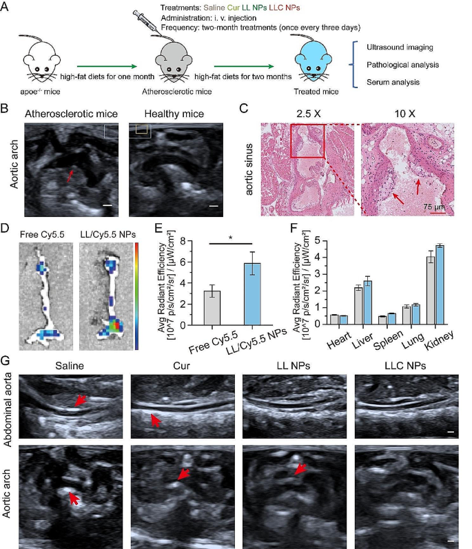
In view of good anti-inflammatory impact, the effectiveness of LLC NPs in countering atherosclerosis was assessed in vivo, following the steps outlined in Fig. 4A. Eight-week-old male apoe−/− mice had been fed with a high-fat eating regimen for a month. Subsequently, the construction of the aortic arch in mice was examined utilizing ultrasound imaging. As proven in Fig. 4B, the inside wall of the aortic arch displayed a pronounced echo, as indicated by the pink arrow, which was the imaging function of arterial atherosclerosis. The formation of froth cells is the primary function of atherosclerotic plaque. Due to this fact, the aortic sinus of mice was eliminated, and H&E staining was performed. In Fig. 4C, there was a noticeable presence of quite a few foam cells within the intima, as proven by the pink body.
As a result of injury to the vascular endothelium and subsequent inflammatory reactions, the permeability of the vascular endothelium at areas with plaques elevated considerably. This alteration facilitates the buildup of nanoparticles (NPs) throughout the plaques. The power of NPs to focus on plaques in vivo was decided by way of fluorescence imaging of extracted tissues, as depicted within the talked about Fig. 4D. 24 h after injection, the aortas from apoe−/− mice handled with Cy5.5-labeled LL NPs confirmed distinguished fluorescence indicators in comparison with these receiving free Cy5.5, indicating NPs had gathered throughout the plaques. Quantitative evaluation of the fluorescence additional highlighted the superior accumulation of Cy5.5-labeled LL NPs within the aorta over free Cy5.5, underscoring the NPs’ outstanding capacity to focus on plaques successfully (Fig. 4E). Moreover, vital fluorescence was noticed in each the liver and kidneys, indicating these organs primarily course of and metabolize NPs (Fig. 4F).
Following the outlined therapy routine (Fig. 4A), ultrasound imaging of the stomach aorta and aortic arch revealed post-treatment adjustments in Fig. 4G. Enhanced echoes, indicating atherosclerotic plaques, had been seen within the saline-treated apoe−/− mice, particularly alongside the stomach aorta’s inside partitions. In distinction, mice handled with NPs displayed much less echo enhancement, suggesting a discount in atherosclerosis, with LLC NPs displaying notable plaque development inhibition. Ultrasound additionally confirmed aortic arch wall thickening in a number of teams, however LLC NPs maintained smoother vascular partitions, highlighting their potential therapeutic advantages in opposition to atherosclerosis.
Imaging analysis of NPs focused remedy for plaque. (A) The therapy plan for atherosclerosis prevention and administration. (B) Ultrasound imaging of aortic arch in early atherosclerotic mice and wholesome mice (Scale bar = 2 mm). (C) H&E staining of aortic sinus in early atherosclerotic mice (Scale bar = 75 μm). (D) Ex vivo fluorescence photographs of the remoted aorta. (E, F) The quantitative common fluorescence depth measurements for the aorta (E) and first organs (F) throughout all experimental teams. Imply ± SD, n = 3, *p < 0.05. (G) Ultrasound photographs of stomach aorta and aortic arch after therapy (Scale bar = 2 mm)
Evaluation of pathological indexes of atherosclerotic plaque
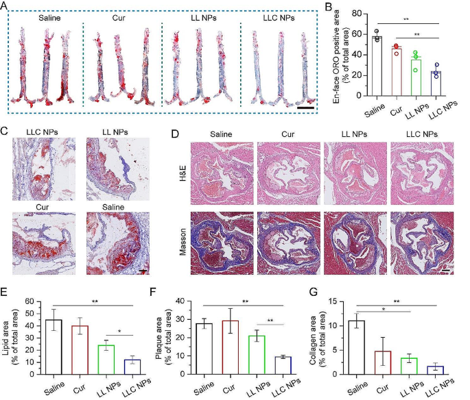
In gentle of optimistic focusing on impact and imaging analysis, evaluation of pathological indexes of atherosclerotic plaque was furtherly performed. Following the completion of therapy, the whole aorta was fastidiously extracted and the Oil Pink O (ORO) staining of aorta was carried out. As proven in Fig. 5A, a big ORO-positive space might be present in remoted aorta of apoe−/− mice handled with saline, and each Cur and LL NPs demonstrated a modest discount in plaque formation, because of the respective anti-inflammation and anti-monocyte adhesion. In distinction, LLC NPs demonstrated outstanding efficacy in combating atherosclerosis, owing to its wonderful capacity to focus on arterial plaques and cascading anti-inflammatory results. The evaluation of ORO-stained areas throughout the aorta (Fig. 5B) revealed that the therapeutic efficacy of LLC NPs drastically surpassed that of free Cur. As well as, sections of aortic sinus had been additionally stained with ORO for evaluation of lipid content material in plaques. As proven in Fig. 5C, the pink space represented the lipids and the lipid content material within the plaques of mice handled with LLC NPs was considerably decrease than that of different teams, which was according to the outcomes of the ORO staining of aorta. Moreover, to investigate the speed of luminal stenosis and the collagen content material inside plaques, the H&E and Masson staining of aortic sinus sections was performed. As proven in Fig. 5D, there have been apparent lipid swimming pools within the plaques of mice within the saline group, however solely foam cells had been discovered on the vascular wall of mice within the therapy group, which indicated that LLC NPs may considerably alleviate the method of atherosclerosis. In addition to, collagen had penetrated into the plaques of mice within the saline group, whereas there was no vital collagen era within the plaques of different therapy teams. We additional quantitatively calculated the luminal stenosis price and collagen content material within the aortic sinus, and located that LLC NPs may considerably cut back the luminal stenosis price and collagen content material within the plaque, additional indicating the superb therapeutic impact of LLC NPs (Fig. 5E-G).
Evaluation of the therapeutic impact of NPs on arterial plaque base on histological staining. (A) Picture of ORO-stained en face aortas (Scale bar = 5 mm). (B) Measurements of areas staining optimistic for ORO in aortas subjected to numerous remedies. Imply ± SD, n = 3, **p < 0.01. (C) Consultant photographs of aortic sinus stained with ORO (Scale bar = 150 μm). (D) Exemplary photographs of sections from the aortic sinus, highlighted by H&E and Masson staining (Scale bar = 250 μm). (E–G) Quantitative proportion of plaque in aortic sinus part samples from ORO (C), H&E and Masson (D) staining. Imply ± SD, n = 3, *p < 0.05, **p < 0.01
Mechanism of anti-atherosclerosis motion and security analysis
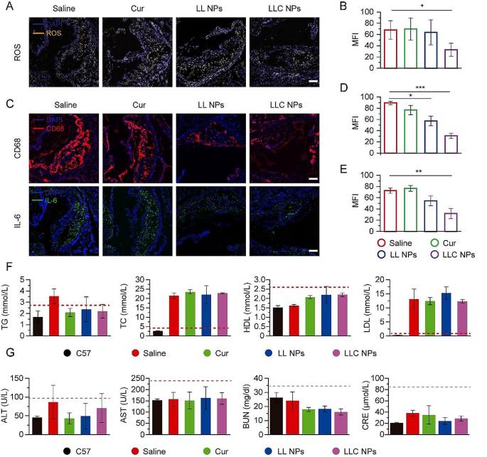
Based mostly on the great anti-atherosclerosis impact of LLC NPs in vivo, we additional analyzed the organic indicators inside plaque to review the therapeutic mechanism. Firstly, the ROS throughout the plaque of aortic sinus was investigated. As proven in Fig. 6A, yellow fluorescence represented areas with excessive expression of ROS, which had been concentrated contained in the plaque. In contrast with the saline group, the yellow fluorescence in plaques of LLC NPs group was considerably weakened, displaying an excellent anti-ROS impact, which was attributed to ROS responsive drug launch and antioxidant exercise of Cur (Fig. 6B). ROS primarily originated from the secretion of macrophages in plaques, so the content material of macrophages in plaques was additionally investigated. In Fig. 6C, pink fluorescence represented macrophages, which had been extensively distributed on the floor and inside plaques in saline group. Free Cur had nearly no therapeutic impact as a consequence of its poor water solubility. The NPs therapy group confirmed low and weak pink fluorescence as a consequence of its inhibition of monocyte adhesion, thereby lowering the content material of macrophages in plaques, and their impact had been considerably higher than that of the saline group (Fig. 6D). Furthermore, IL-6, as a consultant inflammatory issue, was additionally detected. The inexperienced fluorescence represented IL-6, which was additionally distributed contained in the plaque. Per macrophage distribution, IL-6 in plaque was clearly decreased in LLC NPs group (Fig. 6E).
Blood lipid was an necessary index to detect the course of atherosclerosis. Due to this fact, the lipid content material within the blood of mice had been recorded. We discovered that the overall ldl cholesterol (TC) and low density lipoprotein (LDL) ranges within the blood of the atherosclerotic mice had been a lot increased than these of the wholesome mice. Nevertheless, there was no vital distinction in blood lipids indicators, together with TC, triglycerides (TG), LDL, and high-density lipoprotein (HDL), among the many therapy teams (Fig. 6F). The above outcomes indicated that the anti-atherosclerosis mechanism of LLC NPs was primarily associated to the cascade inhibition of ROS and anti-inflammation, and had nothing to do with lipid regulation. For security analysis, liver and kidney operate indicators had been detected in Fig. 6G. There weren’t apparent variations in ALT, AST, BUN and CRE amongst teams. No apparent injury was noticed within the H&E stained sections of the liver, spleen, and kidneys (Fig. S7). In addition to, Physique weight of mice remained constant throughout all teams through the therapy, suggesting that LMWH-LA as drug supply system had an excellent biosafety profile (Fig. S8).
Pathological evaluation and serum evaluation. (A) Fluorescent photographs of aortic sinus stained with dihydroethidium (DHE) and DAPI from atherosclerosis mice (Scale bar = 100 μm). (B) Quantitative evaluation of ROS optimistic space from (A). Imply ± SD, n = 3, *p < 0.05. (C) Sections of aortic sinus stained with antibody to CD68, IL-6 (Scale bar = 100 μm). (D, E) Quantitative evaluation of CD68 optimistic and IL-6 optimistic space from (C). Imply ± SD, n = 3, *p < 0.05, **p < 0.01, ***p < 0.0001. (F) The serum degree of TG, TC, HDL and LDL in all teams. Imply ± SD, n = 4. (G) Indices of hepatic and renal features after therapy in apoe−/− mice. Means ± SD, n = 3

