Synthesis and characterization of CDots
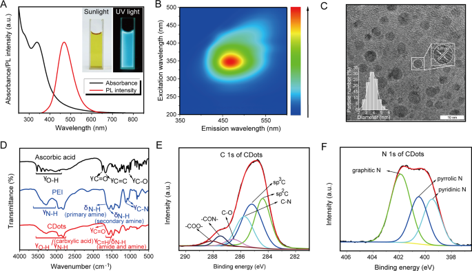
On this examine, carbon dots (CDots) have been synthesized by a one-step microwave method utilizing ascorbic acid and PEI [16]. Beneath daylight, the as-prepared CDots options exhibited a pale yellow, remodeling right into a shiny blue photoluminescence (PL) underneath ultraviolet (UV) radiation (Fig. 1A). The CDots options’ PL spectrum had a single emission peak at roughly 460 nm with a 350 nm excitation wavelength, in distinction to ascorbic acid and PEI which confirmed no PL emission underneath UV mild (Fig. 1B and Determine S1A). Excessive-resolution transmission electron microscopy (HRTEM) revealed that the CDots have been uniform in morphology and had a constant diameter of three.5 nm(Determine 1C). The noticed lattice spacing of 0.21 nm corresponds to the (100) planes of graphene carbon, implying a graphene-like construction [17]. The CDots had a 15.7 mV potential (Determine S1B). Power-dispersive X-ray spectroscopy (EDS) evaluation demonstrated that CDots contained C, N and O components (Determine S1C). The Fourier-transform infrared spectroscopy (FTIR) spectra of ascorbic acid, exhibited C-O stretching vibrations at 1027 cm− 1, C = O stretching vibrations at 1753 cm− 1, and O-H stretching vibrations between 2916 and 3525 cm− 1, suggesting the presence of carboxyl and hydroxyl teams (Fig. 1D). The FTIR spectra of PEI revealed two C-N stretching vibrations at 1120 and 1057 cm− 1 and two N-H bending vibrations at 1662 and 1593 cm− 1, indicative of main and secondary amines (Fig. 1D). The FTIR spectra of those CDots confirmed O-H/N-H stretching vibrations between 2800 and 3600 cm− 1 and a C = O stretching vibration at 1720 cm− 1, indicating the presence of carboxyl and amine teams (Fig. 1D). Moreover, a broad band at 1592 cm− 1 encompassed C = O stretching (amide I band), N-H bending (amide II band), and free amine N-H bending vibrations, ensuing within the overlap of amide I and II bands.
Characterizations of CDots. (A) PL emission spectra (crimson) and UV–vis absorption (black) of CDots. Insets: pictures of CDots answer taken underneath UV mild (proper) and daylight (left), respectively. (B) The excitation-emission map of CDots. (C) HRTEM picture of CDots. (D) FTIR spectra of CDots (crimson), PEI (blue) and ascorbic acid (black). (E–F) Excessive-resolution XPS spectra of CDots: E is C 1s, F is N 1s
These outcomes confirmed that the formation of CDots was on account of dehydration condensation and additional carbonization between ascorbic acid and PEI, with a large number of purposeful teams current within the CDots, as additional evidenced by X-ray photoelectron spectroscopy (XPS) characterization. The C 1s spectrum of ascorbic acid revealed 4 peaks at 284.7, 285.7, 286.6, and 288.4 eV, equivalent to sp2 C, sp3 C, C-O, and -COO- teams. The N 1s spectrum of PEI, centered at 399.9 eV (Determine S1D), was related to amine (-NH-) teams. In distinction, the C 1s spectrum of the CDots includes six peaks at 284.6, 285.4, 285.9, 286.7, 287.7, and 288.3 eV (Fig. 1E), representing sp2 C, sp3 C, C-N, C-O, -CON-, and -COO- teams. The presence of -CON- was because of the amidation response between ascorbic acid and PEI, whereas the sp2 C species indicated the existence of extremely conjugated sp2 domains within the CDots, in keeping with HRTEM outcomes (graphene-like buildings) (Fig. 1C). Moreover, the N 1s spectrum of the CDots confirmed three peaks at 399.3, 400.5, and 401.9 eV (Fig. 1F), attributable to pyridinic, pyrrolic, and graphitic nitrogen, indicating the presence of heterocyclic bases. In abstract, these outcomes revealed the formation technique of the CDots and the presence of purposeful teams inside them.
CDots exhibit glorious biocompatibility
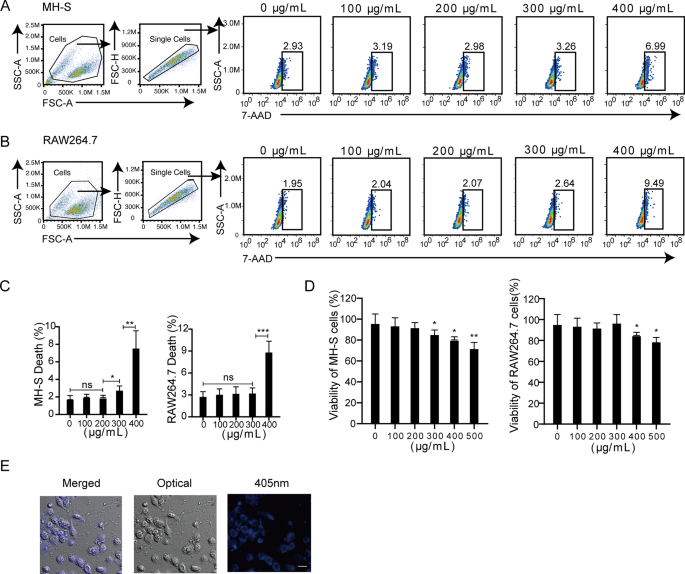
Biocompatibility is an important prerequisite for CDots purposes. On this regard, we evaluated the results of CDots on cell viability and located that CDots exhibited no vital cytotoxic results on each mouse alveolar macrophage MH-S cells and mouse macrophage RAW264.7 cells at concentrations under 200 µg/mL. Nevertheless, vital cytotoxic results have been noticed at concentrations of 300 and 400 µg/mL(Fig. 2A-C). In step with the circulation cytometric outcomes, CCK-8 assays corroborated {that a} CDots focus of 200 µg/mL didn’t considerably influence the cell viability of each MH-S and RAW264.7 cells (Fig. 2D). Subsequently, for subsequent experiments, a CDots answer at a focus of 200 µg/mL was chosen. Macrophages have an intrinsic phagocytic perform, which may swallow exogenous substances into the host cells [18]. Based mostly on the blue fluorescence property of CDots, we traced the intracellular presence of CDots in MH-S cells utilizing laser confocal scanning microscopy (CLSM). CDots might enter and persist in mouse macrophage cells for no less than 24 h (Fig. 2E). Thus, these outcomes reveal the superb biocompatibility of CDots with cells.
Cytotoxicity assays and imaging of CDots in mouse macrophages. (A–C) Consultant gating Methods (A–B) and quantification of cell demise (C) by 7-AAD staining. MH-S and RAW264.7 cells have been handled with CDots at indicated concentrations. (D) Quantification of CCK-8 assays. MH-S and RAW264.7 cells have been handled with CDots at indicated concentrations. (E) Cell imaging of CDots-treated MH-S cells, that are merged picture, optical picture, and CLSM (405 nm) picture from left to proper. Statistical significance was analyzed by the two-tailed Pupil’s t-test. Information are introduced as imply ± SD from three impartial experiments. *p < 0.05, **p < 0.01, ***p < 0.001. Scale bar = 10 μm
CDots exert a therapeutic impact on bacterial pneumonia in mice
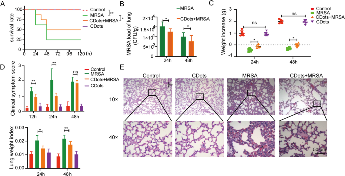
The toxicity of CDots was first investigated in mice by intravenous injection of CDots. Hematoxylin and eosin (H&E) staining revealed that 48 h after administering CDots, there was no vital injury noticed in varied organs in comparison with the management group (Determine S2), demonstrating the biocompatibility of CDots and their suitability for additional in vivo research. To evaluate the therapeutic efficacy of CDots, we established MRSA or Okay. pneumoniae-induced pneumonia mannequin in mice, who have been handled with an intravenous injection of CDots 6 h post-infection. In comparison with the bacterial an infection group, CDots therapy considerably enhanced the survival charges of mice, and markedly diminished lung bacterial masses (Fig. 3A-B, Determine S3A-B), weight reduction, medical scores, and lung indices, amongst different medical indicators (Fig. 3C-D, Determine S3C-D). Moreover, H&E staining of lung tissues at 24 h post-infection with MRSA and Okay. pneumoniae revealed in depth neutrophil infiltration, tissue degeneration, necrosis, ample fibrin exudation, and erythrocyte extravasation within the alveoli. Remarkably, there have been nearly no crimson blood cells noticed partaking in regular fuel trade throughout the alveoli. CDots therapy, nonetheless, considerably mitigated the aforementioned pathological injury (Fig. 3E, Determine S3E). Moreover, to research the influence of the ratio of ascorbic acid to PEI on therapeutic efficacy, we synthesized further batches of CDots by growing the quantity of ascorbic acid (leading to a 0.25:1 ratio of PEI to ascorbic acid) or the quantity of PEI (leading to a 1:1 ratio of PEI to ascorbic acid) (Determine S4A). These CDots with completely different proportions exhibited related UV-visible spectra (Determine S4B), and therapeutic efficacy (Determine S4C-J). Collectively, these in vivo outcomes show that CDots exerted a therapeutic impact on MRSA and Okay. pneumoniae-induced bacterial pneumonia in mice.
Analysis of the therapeutic impact of CDots on bacterial pneumonia in mice. (A) The survival price of the mice in numerous therapy teams (n = 8). (B) Bacterial masses in murine lung homogenate (n = 6). (C–D) Scientific index of the mice in numerous therapy teams, together with weight improve (C), medical symptom rating (D), and lung weight index (D) (n = 6). (D) H&E staining outcomes of lung pathological sections in numerous teams. Statistical significance was analyzed by the two-tailed Pupil’s t-test. Information are introduced as imply ± SD from three impartial experiments. *p < 0.05, **p < 0.01, ***p < 0.001. Scale bar = 50 μm
CDots Promote the M1 Polarization of Macrophages to Increase Their Cell Vitality and Phagocytic Antimicrobial Results.
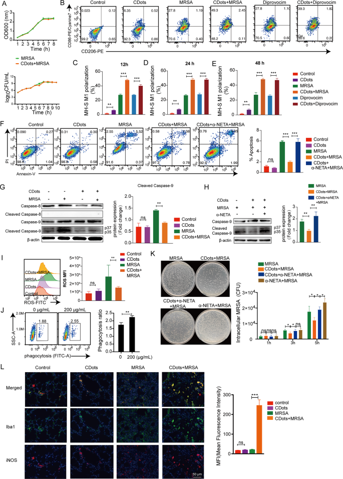
Within the realm of resisting pathogenic microbial infections, CDots have been primarily designed to straight goal the micro organism to elicit antimicrobial results [19]. Subsequently, the as-prepared CDots have been first launched straight into the bacterial tradition medium of MRSA and Okay. Pneumoniae to look at their direct bactericidal capabilities. Surprisingly, CDots exhibited no vital inhibitory impact on the proliferation of each MRSA and Okay. Pneumoniae (Fig. 4A, Determine S5A).
Results of CDots on macrophage polarization, cell vitality, phagocytosis, and sterilization. (A) The lengthy curve measurement and colony rely have been measured for MRSA with or with out CDots. (B-E) Consultant gating methods for CD206 and CD86 expression in MH-S cells and quantification of CD86+ macrophages after indicated therapies. (F) Consultant circulation cytometric plots and quantification of cell apoptosis. (G) Western blot outcomes of caspase-8, cleaved caspase-8; caspase-9 and cleaved caspase-9 of MH-S cells handled as indicated. (H) Western blot outcomes of caspase-9 and cleaved caspase-9 of MH-S cells handled as indicated. (I) Circulation cytometry evaluation of intracellular ROS modifications in MH-S cells. (J) Circulation cytometric evaluation of the phagocytosis of MH-S cells with or with out CDots therapies. (Okay) Consultant pictures of micro organism after 5 hours’ an infection and the quantification of bacterial colonies with indicated therapies. (L) The expression of iNOS+ macrophages in murine lung sections was evaluated by immunohistochemistry (Iba1+: inexperienced; iNOS+: crimson; DAN: blue). The histogram confirmed the typical fluorescence depth of Iba 1 and iNOS co-localization. Statistical significance was analyzed by the two-tailed Pupil’s t-test. Information are introduced as imply ± SD from three impartial experiments. *p < 0.05, **p < 0.01, ***p < 0.001
Following this, we additional explored whether or not CDots exert their antibacterial results by modulating macrophage perform. Circulation cytometric evaluation revealed that, with or with out MRSA, Okay. pneumoniae, TLR2 agonist (Diprovocim) and TLR4 agonist (RS 09) stimulation, CDots considerably enhanced the proportion of CD86+ M1-type macrophages (Fig. 4B-E, Determine S5B-F). Extending the incubation time of CDots had no vital results on the proportion of macrophage polarization (Fig. 4C-E, Determine S5D-F). As well as, cell apoptosis detection demonstrated that CDots considerably diminished macrophage apoptosis following an infection with MRSA and Okay. Pneumoniae (Fig. 4F, Determine S5G-H). Subsequent, we explored the pathways by which CDots particularly affected apoptosis, and located CDots might lower MRSA-induced apoptosis by decreasing the expression of cleaved caspase-9 (Fig. 4G). Nevertheless, the M1 polarization inhibitor (α-NETA) markedly mitigated this protecting impact by cleaved caspase-9 (Fig. 4H), suggesting that CDots can shield macrophages from apoptosis throughout MRSA an infection by selling M1 polarization. On the identical time, CDots therapy might scavenge extra ROS (Fig. 4I, Determine S5I). Furthermore, CDots enhanced the phagocytic capability of mouse alveolar macrophages (Fig. 4J, Determine S5J ). CDots addition might remarkably scale back the intracellular bacterial load in macrophages whereas this impact was negated when an M1 polarization inhibitor was added (Fig. 4Okay, Determine S5Okay). Constant outcomes have been obtained when utilizing mouse macrophages RAW264.7 (Determine S6). Moreover, the M1 polarization of macrophages in murine lungs following CDots administration was investigated. In step with the outcomes of in vitro experiments, CDots might considerably improve the frequencies of lung iNOS+ M1-macrophages in MRSA an infection (Fig. 4L). Taken collectively, these outcomes demonstrated that CDots might promote the M1 polarization of macrophages to reinforce their cell vitality and phagocytic bactericidal exercise.
CDots promote macrophage M1 polarization by inhibiting the activation of PI3K/AKT/mTOR signaling pathway
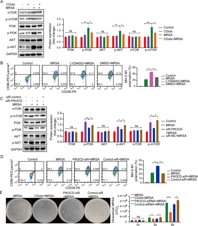
Quite a few research have established a powerful affiliation between the M1 polarization course of and the activation of the PI3K/AKT/mTOR signaling pathway [20]. We additional discover the connection between CDots-promoted M1 polarization and the PI3K pathway. After MRSA or Okay. pneumoniae an infection, the protein expression ranges of p-PI3K, p-AKT, and p-mTOR considerably elevated, whereas CDots notably diminished the phosphorylation ranges of those proteins (Fig. 5A, Determine S7A). Subsequently, the PI3K signaling pathway inhibitor LY294002 was used to look at its results on macrophage polarization. Each the PI3K pathway inhibitor and CDots considerably promoted the macrophage M1 polarization, suggesting that CDots might promote M1 polarization of macrophage by inhibiting the activation of the PI3K signaling pathway (Fig. 5B, Determine S7B). PIK3CD, the catalytic subunit of PI3K, performs a vital position in activating the PI3K signaling pathway [21]. To determine whether or not CDots exert their results by focusing on PIK3CD, we investigated the results of PIK3CD knockdown on macrophages. In comparison with the Management-SiR group, the protein ranges of p-mTOR, p-PI3K, and p-AKT have been considerably diminished within the siRNA-PIK3CD group for each MH-S and RAW264.7 cells (Fig. 5C, Determine S7C and Determine S8A). Moreover, PIK3CD knockdown elevated the proportion of CD86+ M1 macrophages (Fig. 5D, Determine S7D) and enhanced the bactericidal exercise of macrophages, just like these noticed with CDots (Fig. 5E, Determine S7E and Determine S8B). Thus, these information prompt that CDots might goal PIK3CD to inhibit the activation of the PI3K signaling pathway, subsequently selling M1 polarization.
CDots argument M1 polarization by way of inhibiting the PI3K/AKT/mTOR signaling pathway. (A, C) Western blot outcomes of PI3K, p-PI3K, AKT, p-AKT, mTOR, and p-mTOR, of MH-S cells handled as indicated. (B, D) Circulation cytometric evaluation of CD206 and CD86 expression in MH-S cells with indicated therapies. (E) Consultant pictures of micro organism after 5 hours’ an infection and the colonies of micro organism with completely different therapies. Statistical significance was analyzed by the two-tailed Pupil’s t-test. Information are introduced as imply ± SD from three impartial experiments. *p < 0.05, **p < 0.01, ***p < 0.001, ****p < 0.0001
CDots can straight bind to PIK3CD
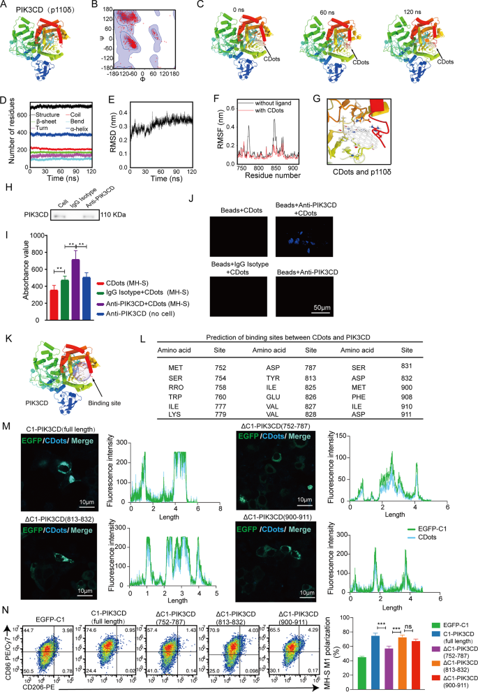
CDots have been demonstrated to modulate macrophage perform by the PI3K signaling pathway, we subsequent explored the potential interplay mechanisms between PIK3CD and CDots. The zeta potential of CDots is optimistic, whereas PIK3CD carries a detrimental cost, offering a foundation for electrostatic attraction between the 2. Based mostly on this construction, we hypothesize that CDots work together with PIK3CD to type steady complexes, exerting organic results. Molecular dynamics simulations have been first carried out to evaluate the steadiness of the complexes. A simplified mannequin of CDots, represented by a graphene-like fragment, was connected to the ligand molecule, and the PIK3CD construction was constructed utilizing the homology modeling technique [22], based mostly on buildings obtained from the RCSB Protein Information Financial institution (PDB ID: 2WXF) (Fig. 6A). The standard of the homology mannequin was validated, with over 99% of PIK3CD residues positioned throughout the allowed areas of the Ramachandran plot (Fig. 6B). Molecular dynamics simulations have been carried out for 120 ns, and the simulation trajectory was captured by snapshot pictures (Fig. 6C and Film S1). All through all the simulation length, the CDots remained sure to the CDots-PIK3CD advanced, indicating robust binding affinity. The secondary buildings of PIK3CD, together with α-turns, helices, β-bridges, and β-sheets, remained almost unchanged all through the simulation (Fig. 6D). The structural stability of the advanced was additional confirmed by the foundation imply sq. deviation (RMSD), which quickly stabilized with minimal fluctuations (Fig. 6E). These outcomes recommend that CDots don’t adversely have an effect on the structural integrity of PIK3CD. Following this, we assessed the foundation imply sq. fluctuation (RMSF) of every amino acid residue in PIK3CD to guage their positional variance. Earlier than inspecting the RMSF throughout the advanced, a 120 ns molecular dynamics simulation of the PIK3CD, within the absence of any ligand, was carried out. The RMSF values obtained for PIK3CD have been used as a reference for comparability. The RMSF confirmed that the amino acid residues of PIK3CD within the CDots-PIK3CD advanced have been extra restrained in comparison with the free PIK3CD, indicating robust interactions between PIK3CD and CDots (Fig. 6F and G).
Molecular dynamics simulation and verification of interplay between CDots and PIK3CD. (A) Three-dimensional construction of PIK3CD. Secondary structural components are depicted as tube helices. (B) Ramachandran plot of PIK3CD. (C) The buildings of CDots binding to PIK3CD, that are extracted from the molecular dynamics simulation trajectory at 0, 60, and 120 ns, respectively. (D) The variation within the numbers of residues inside completely different secondary buildings of PIK3CD all through the simulation. (E) RMSD of the Cα atoms of PIK3CD in PIK3CD-CDots advanced towards time. (F) RMSF values of PIK3CD and within the PIK3CD-CDots advanced, respectively. (G) 3D diagram of the interplay between CDots and PIK3CD. Amino acid residues concerned within the binding of CDots are drawn as sticks. (H) The Immunoprecipitation experiment of PIK3CD. Western blotting confirmed that there was a transparent PIK3CD band after Anti-PIK3CD IP, which proved that the PIK3CD antibody had sure PIK3CD. (I) The fluorescence information of the immunoprecipitation advanced after eradicating the beads have been collected with a VICTOR X5 Multilabel Plate Reader. CDots (MH-S) represents immunoprecipitation obtained with out antibody; IgG Isotype + CDots (MH-S) represents immunoprecipitation obtained utilizing isotype management; Anti-PIK3CD + CDots (MH-S) represents immunoprecipitation obtained utilizing PIK3CD antibody; Anti-PIK3CD + CDots (no cell) represents immunoprecipitation obtained utilizing PIK3CD antibody with out cells. (J) The fluorescence of immunoprecipitation complexes with beads was detected by confocal pictures. *p < 0.05, **p < 0.01. Scale bar = 50 μm. (Okay)Molecular dynamics simulation trajectory prediction of the structural area of CDots binding to PIK3CD. (L) Desk of amino acid residues in PIK3CD with potential interactions with CDots obtained from molecular dynamics simulations. (M) Laser confocal microscopy shows the binding of CDots to PIK3CD and its deletion types, the place inexperienced fluorescence represents the expression of PIK3CD and its deletion types, and blue fluorescence represents CDots. The dimensions bars = 10 µ m /20 µ m, respectively. Histogram represents fluorescence depth evaluation of EGFP-C1 (inexperienced line) and CDots (blue line) in 293T cells. (N) The consultant circulation cytometry plots present the M1polarization stage in MH-S cells after CDots bind to PIK3CD and its deletion
Moreover, the interplay between CDots and PIK3CD inside MH-S cells was validated utilizing adjusted immunoprecipitation mixed with laser confocal know-how. Following the therapy, PIK3CD was precipitated utilizing PIK3CD antibody or isotype antibody connected to magnetic beads, with a management group arrange for cell an infection with out immunoprecipitation. After eluting the immunoprecipitated advanced and gathering the liquid, Western blot evaluation revealed a definite PIK3CD band within the PIK3CD antibody (anti-PIK3CD) group in comparison with the Isotype IgG group, indicating profitable binding of PIK3CD molecules by the antibody (Fig. 6H). Using the fluorescent properties of CDots, full-wavelength enzyme-linked immunosorbent assay (ELISA) detected considerably greater values within the Anti-PIK3CD group in comparison with the Isotype IgG group (Fig. 6I), confirming the presence of CDots within the PIK3CD sure. Additional laser confocal evaluation confirmed a major improve in blue mild within the Anti-PIK3CD group in comparison with the isotype management group (Fig. 6J), validating the interplay between CDots and PIK3CD. Lastly, the binding domains of CDots and PIK3CD have been investigated and located that the important thing purposeful area of PIK3CD was positioned throughout the amino acid residues 752 to 911 by molecular dynamics simulations (Fig. 6Okay-L). To additional establish potential binding domains between CDots and PIK3CD, we truncated amino acids 752–787, 813–832, and 900–911 of PIK3CD, respectively, and constructed EGFP-C1 vectors with full-length and three varieties of PIK3CD amino acid sequence deletions. The outcomes confirmed that when the 752–787 amino acid residues of PIK3CD have been deleted, the binding depth of CDots to PIK3CD was weaker, and macrophages exhibited decrease M1 polarization ranges in contrast with full-length and different deletions (Fig. 6M-N). Thus, these information prompt that CDots might straight bind to PIK3CD and the important area for the binding was positioned in amino acids 752–787 of PIK3CD.

