Design precept of the electrochemical biosensor
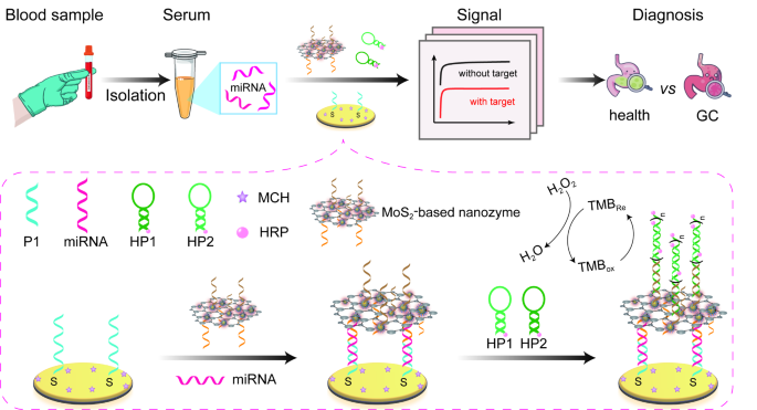
As illustrated in Scheme 1, P1 was firstly assembled on electrode floor by way of Au-S bond. After passivating the electrode floor with MCH, goal miR-19b-3p and P2-HCR-I/MoS2-Au@Pt nanoprobes had been launched to assemble P2-HCR-I/MoS2-Au@Pt/miR-19b-3p/P1/Au because of the formation of P1-miR-19b-3p-P2 sandwiched construction. Then, the assembled HCR-I triggered the incidence of HCR response within the presence of HRP-labelled each HP1 and HP2. Because of this, a lot of HRP had been assembled on the floor of electrode floor with the incidence of HCR response. Due to this fact, the assembled MoS2-Au@Pt nanozymes and HRP can effectively co-catalyze H2O2-TMB response to generate excellent electrochemical sign, reaching the aim of sign amplification. On this examine, a triple sign amplification technique was designed for miR-19b-3p detection. MoS2-Au@Pt nanocomposites aren’t solely used as nanoprobes to load P2 and HCR-I for recognizing and capturing miR-19b-3p, but additionally are employed as nanozymes to catalyze H2O2-TMB response. At this second, this biosensor achieved the primary sign amplification. Within the presence of HP1 and HP2, a typical HCR response was initiated by HCR-I. Accompanied by HCR response, a super-long DNA double-stranded construction labelled with HRP was obtained on electrode floor, ensuing within the second sign amplification. The launched HRP catalyze H2O2-TMB response to attain the third sign amplification. Coupled with the triple sign amplification, this proposed biosensor can qualitatively and quantitatively detect miR-19b-3p with passable outcomes.
Characterization of MoS2, MoS2-AuNPs and MoS2-Au@Pt
Transmission electron microscope (TEM) was used to characterize the morphologies of as-prepared MoS2, MoS2-AuNPs and MoS2-Au@Pt nanomaterials. As proven in Fig. S1, gold nanoparticles (AuNPs) with a mean diameter of 8.1 ± 0.7 nm had been distributed on the floor of MoS2 nanosheets, forming MoS2-AuNPs nanocomposites. Subsequently, MoS2-AuNPs nanocomposites had been used as nanoseeds to arrange MoS2-Au@Pt nanocomposites. Clearly, 20.1 ± 0.5 nm core-shell Au@Pt bimetallic nanoparticles had been supported on MoS2 floor, proving that the profitable synthesis of MoS2-Au@Pt nanocomposites.
Zeta potential evaluation was utilized to guage the costs of the nanocomposite suspensions. As noticed in Fig. S2, the zeta potential of MoS2 nanosheets was − 22.2 mV. In contrast with MoS2 nanosheets, the zeta potential of MoS2-AuNPs nanocomposites was positively shifted to -13.4 mV, suggesting that the profitable ornament of AuNPs [31, 32]. When Au@Pt bimetallic nanoparticles supported on the floor of MoS2 nanosheets, the zeta potential elevated to 11.3 mV, which is ascribed to the positively charged surfactant adsorbed on the nanomaterials floor [33].
Characterization of P2-HCR-I/MoS2-Au@Pt nanoprobes
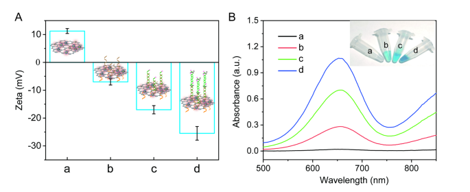
Zeta potential was used to confirm the preparation of P2-HCR-I/MoS2-Au@Pt nanoprobes and the incidence of HCR response. As noticed in Fig. 1A, the zeta potential of MoS2-Au@Pt, P2-HCR-I/MoS2-Au@Pt, HP1/P2-HCR-I/MoS2-Au@Pt and HP1-HP2/P2-HCR-I/MoS2-Au@Pt had been 11.2 mV, -7.0 mV, -17.0 mV and − 25.5 mV, respectively, suggesting that the profitable preparation of nanoprobes and the incidence of HCR response on nanocomposites’ floor. The reason being ascribed to the negatively charged DNA had been assembled on the floor of MoS2-Au@Pt nanocomposites. The extra DNA assembled on nanocomposites’ floor, the zeta potential shifted extra negatively.
Furthermore, the catalytic actions of MoS2-Au@Pt nanoprobes and nanoprobe-assisted sign amplification system had been characterised through the use of H2O2-TMB response system (Fig. 1B). No absorption peak of H2O2-TMB answer was obtained, suggesting that no catalytic response was occurred (curve a). An apparent absorption peak at 652 nm and a blue answer had been noticed when MoS2-Au@Pt nanocomposites was added into H2O2-TMB answer (Fig. S3). This phenomenon effectively proves that MoS2-Au@Pt nanocomposite has a peroxidase-like enzyme exercise to catalyze H2O2-TMB response. As soon as P2 and HCR-I had been co-assembled on the floor of MoS2-Au@Pt nanocomposites to type P2-HCR-I/MoS2-Au@Pt nanoprobes, the absorption peak depth of nanoprobes decreased about 38.3% in contrast with that of MoS2-Au@Pt nanocomposites, indicating that P2 and HCR-I partly blocked the catalytic websites of MoS2-Au@Pt nanocomposites (Fig. S3, curve b in Fig. 1B). To acquire excessive detection efficiency, each HP1 and HP2 labelled with HRP had been launched to enhance the catalytic capacity and attain a number of sign amplification. When HP1 was hybridized on the floor of nanoprobes, response answer grew to become bluer and the absorption peak depth elevated to 0.7 (curve c). The very best absorption peak depth (1.0) and the bluest answer color had been noticed when each HP1 and HP2 had been added into the response technique (curve d), indicating that HCR response was efficiently occurred on nanocomposites’ floor. The biggest response was ascribed to the synergistic impact of MoS2-Au@Pt nanozymes and HRP.
(A) The zeta potentials of (a) MoS2-Au@Pt, (b) P2-HCR-I/MoS2-Au@Pt, (c) HP1/P2-HCR-I/MoS2-Au@Pt and (d) HP1-HP2/P2-HCR-I/MoS2-Au@Pt. (B) UV-visible absorption spectra of (a) H2O2-TMB, (b) H2O2-TMB + P2-HCR-I/MoS2-Au@Pt, (c) H2O2-TMB + HP1/P2-HCR-I/MoS2-Au@Pt and (d) H2O2-TMB + HP1-HP2/P2-HCR-I/MoS2-Au@Pt. Inset: the corresponding {photograph} of various merchandise in H2O2-TMB answer
Characterization of preparation strategy of the electrochemical biosensor
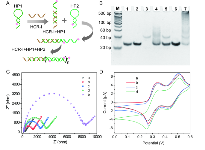
As proven in Fig. 2A, the initiator DNA (HCR-I) can set off the self-assembly of HP1 and HP2 to generate lengthy DNA double-strands (dsDNA). The HCR product was verified by 8% native polyacrylamide gel electrophoresis (Fig. 2B). Clearly, a single band was noticed from lane 1 to lane 3, which is equivalent to HP1, HP2 and HCR-I, respectively. As expectedly, HCR-I can open the hairpin construction of HP1 to type dsDNA construction by way of hybridization response, producing a brand new band in lane 4. In the meantime, two bands had been obtained in lane 5, suggesting that HCR-I doesn’t hybridize with HP2. Furthermore, HP1 and HP2 coexist within the response system with out the set off of HCR-I (lane 6). Within the presence of HCR-I, HCR-I initiates HCR response to provide lengthy dsDNA. Correspondingly, ladder bands appeared in lane 7.
Electrochemical impedance spectroscopy (EIS) and cyclic voltammetry (CV) had been carried out to observe the fabrication strategy of this electrochemical biosensor. Usually, the electron switch resistance (Rct) suggests the interfacial impedance of various modified electrodes. If the damaging costs of P1 and miR-19b-3p had been assembled on the naked gold electrode floor (curve a, Fig. 2C), the Rct of P1/Au (curve b, Fig. 2C) and miR-19b-3p/P1/Au (curve c, Fig. 2C) elevated to 1407.4 Ω and 2426.2 Ω, respectively. With the meeting of P2-HCR-I/MoS2-Au@Pt nanoprobes, the Rct worth was elevated to 3207.5 Ω (curve d, Fig. 2C). As soon as the incidence of HCR response, the launched HP1 and HP2 triggered an excellent enhance within the Rct worth of HP1-HP2/P2-HCR-I/MoS2-Au@Pt/miR-19b-3p/P1/Au (8483.4 Ω, curve e, Fig. 2C). These enhance of Rct values proved the profitable preparation of the designed electrochemical biosensor. Electrochemical behaviors of various preparation processes had been additionally characterised by CV in [Fe(CN)6]4−/3−answer (Fig. S4). Notably, CV outcomes had been in settlement with EIS outcomes, furtherly proving that the designed biosensor have been efficiently constructed.
The electrochemical behaviour of this biosensor was additionally studied within the TMB and H2O2 combination answer. As illustrated in Fig. 2D, two pairs of redox peaks had been noticed at P1/Au, which was ascribed to the standard two electron redox response of TMB within the presence of H2O2 (curve a). After the profitable meeting of MoS2-based nanoprobes, just a few enhance within the peak present at P2-HCR-I/MoS2-Au@Pt/miR-19b-3p/P1/Au was noticed (curve b). The assembled MoS2-based nanoprobes have electrocatalytic exercise, which might catalyze the response of H2O2-TMB. When HRP-labelled HP1 and HRP-labelled HP2 had been launched on the electrode floor, an apparent amplified electrochemical responses had been obtained at HP1/P2-HCR-I/MoS2-Au@Pt/miR-19b-3p/P1/Au (curve c) and HP1-HP2/P2-HCR-I/MoS2-Au@Pt/miR-19b-3p/P1/Au (curve d), respectively. The height currents had been drastically trusted the quantity of HRP, which might effectively catalyze H2O2-TMB response to generate an excellent electrochemical sign. All experimental information prompt that the synergistic impact of MoS2-based nanoprobes, HCR response and HRP enzyme can amplify electrochemical sign, which is helpful for hint goal molecules detection.
(A) Schematic diagram of HCR course of. (B) Totally different merchandise had been characterised by gel electrophoresis. From lane M to lane 7 is 20 bp DNA ladder marker, HP1, HP2, HCR-I, HCR-I + HP1, HP2 + HCR-I, HP1 + HP2, HCR-I + HP1 + HP2, respectively. (C) EIS curves of (a) naked Au, (b) P1/Au, (c) miR-19b-3p/P1/Au, (d) P2-HCR-I/MoS2-Au@Pt/miR-19b-3p/P1/Au, (e) HP1-HP2/P2-HCR-I/MoS2-Au@Pt/miR-19b-3p/P1/Au in 5 mM Okay3[Fe(CN)6]/Okay4[Fe(CN)6] answer containing 0.1 M KCl. (D) CV curves of (a) P1/Au, (b) P2-HCR-I/MoS2-Au@Pt/miR-19b-3p/P1/Au, (c) HP1/P2-HCR-I/MoS2-Au@Pt/miR-19b-3p/P1/Au, (d) HP1-HP2/P2-HCR-I/MoS2-Au@Pt/miR-19b-3p/P1/Au in H2O2-TMB answer
Feasibility of sign amplification technique
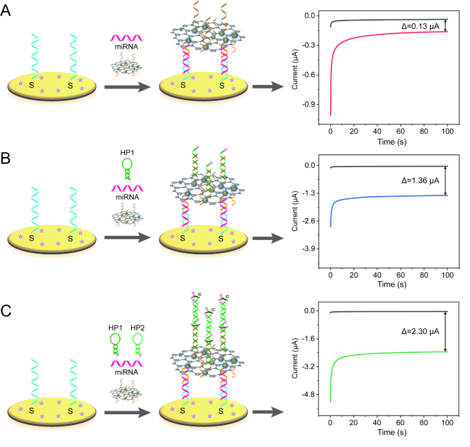
The sign amplification impact of this biosensor was investigated. As talked about in Scheme 1, MoS2-based nanozymes, HCR and HRP had been used to amplify electrochemical sign. For effectively notice the sign amplification impact, two management teams included single sign amplification (MoS2-based nanoprobe) and double sign amplification (MoS2-based nanoprobe and HRP) had been designed for comparability (Fig. 3). For a similar focus of miR-19b-3p detection (100 pM), the height currents variations (ΔI = ImiRNA − Ino miRNA) of single sign amplification technique had been 0.13 µA (Fig. 3A). Against this, the ΔI of double sign amplification was reached to 1.36 µA, which is sort of 10.46 occasions than that amplified by single sign amplification (Fig. 3B). For triple sign amplification technique, the ΔI was about 2.30 µA, which is about 17.69 occasions and 1.69 occasions than that of single sign amplification and double sign amplification technique, respectively, suggesting that the triple sign amplification has one of the best electrochemical response (Fig. 3C).
Electrochemical biosensors with completely different sign amplification methods (A) single sign amplification (MoS2-based nanoprobes), (B) double sign amplification (MoS2-based probes + HRP catalytic response), (C) triple sign amplification (MoS2-based nanoprobe + HCR response + HRP catalytic response) for 100 pM miR-19b-3p detection in H2O2-TMB answer. The preliminary potential was 150 mV and the runtime was 100 s
Analytical efficiency of the electrochemical biosensor
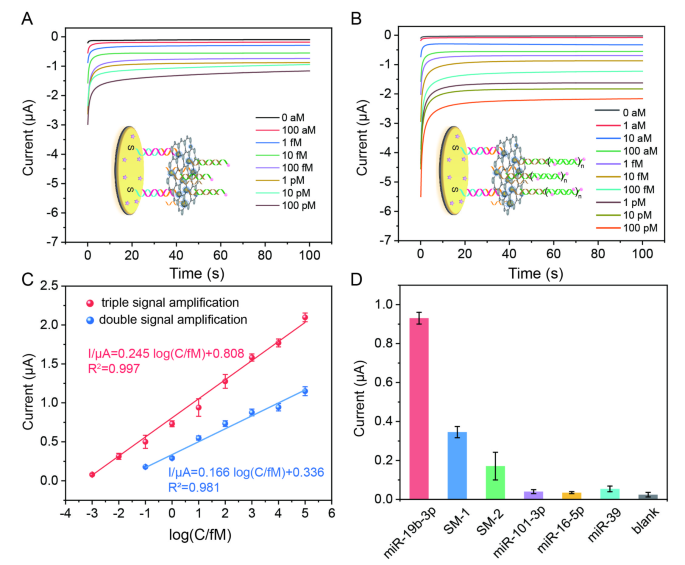
To acquire the optimum efficiency of the electrochemical biosensor, the experimental situations had been optimized. As displayed in Fig. S5A, the optimum incubation time of MoS2-Au@Pt-based nanoprobes and the response time of HCR had been chosen as 80 min and 80 min, respectively. Beneath the optimum situations, the analytical efficiency of electrochemical biosensors coupled with double sign amplification (MoS2-based nanoprobes + HRP) and triple sign amplification (MoS2-based nanoprobe + HCR + HRP) for miR-19b-3p detection was examined through the use of amperometric technique, respectively. As proven in Fig. 4A and B, the currents of the biosensors amplified by double sign amplification and triple sign amplification had been growing with the addition of miR-19b-3p, respectively. From the amperometric curves, two linear relationships between the present worth on the a hundredth second and the logarithm of miR-19b-3p concentrations had been obtained with equations of (:I/{upmu:}textual content{{rm:A}}=0.245text{log}left(C/fMright)+0.808) (R2 = 0.997) and (:I/{upmu:}textual content{{rm:A}}=0.166text{log}left(C/fMright)+0.336) (R2 = 0.981) for triple sign amplification- and double sign amplification-based biosensors, respectively (Fig. 4C). Clearly, the triple sign amplification-based biosensor has wider dynamic vary (1 aM-100 pM vs. 100 aM-100 pM), bigger electrochemical response (0.50 µΑ vs. 0.18 µΑ for 100 aM miR-19b-3p detection) and decrease detection restrict (0.7 aM vs. 37.4 aM) than the double sign amplification-based biosensor. The analytical efficiency of this triple sign amplification-based biosensor is healthier than some revealed works for miRNA detection (Desk S2). The selectivity of this triple sign amplification-based biosensor was examined. As proven in Fig. 4D, the present of this biosensor for 10 fM miR-19b-3p detection (0.93 µΑ) is about 2.7 occasions, 5.4 occasions, 23.3 occasions, 26.6 occasions, 17.2 occasions than this biosensor for 10 pM two single-base mismatched miR-19b-3p (SM-1, SM-2), miR-101-3p, miR-16-5p, miR-39 detection, respectively, suggesting that this biosensor can effectively distinguish goal miR-19b-3p from even a single-base mismatched microRNA sequence. The reproducibility of the biosensor was studied through the use of ten impartial biosensors. As proven in Fig. S6, the relative commonplace deviation (RSD) of intra- and inter-assays had been 3.89% and 1.70%, respectively, suggesting that the biosensor has good reproducibility. The storage stability of this biosensor was studied by storing the developed electrodes at 4 °C within the fridge. Experimental outcomes confirmed that the response present of this biosensor was about 92.8% of the preliminary sign response after 8 days storage, indicating that the biosensor has wonderful stability (Fig. S7).
(A) I-t responses of the electrochemical biosensor coupled with double sign amplification methods for various concentrations of miR-19b-3p detection in H2O2-TMB answer. (B) I-t responses of the triple sign amplification-based biosensor for various concentrations of miR-19b-3p detection in H2O2-TMB answer. The preliminary potential was 150 mV and the runtime was 100 s. (C) The linear relationships between the present worth and the logarithm of miR-19b-3p concentrations. (D) The selectivity of the biosensor for the detection of 10 fM miR-19b-3p, 10 pM SM-1, 10 pM SM-2, 10 pM miR-101-3p, 10 pM miR-16-5p, and 10 pM miR-39, respectively. The illustrated error bars signify the usual deviations of not less than three impartial measurements (n = 3)
Evaluation of miR-19b-3p in scientific samples
At first, simulated serum pattern was used to guage the sensible analytical capacity of this biosensor. Usually, completely different concentrations (100 aM, 1 fM, 10 fM, 100 fM and 1 pM) of miR-19b-3p had been added in 1% human serum to simulate the actual samples. As displayed in Desk 1, the recoveries and relative commonplace deviation (RSD) of this biosensor are about 95.2-102.5% and a couple of.1-5.8%, respectively, proving that the proposed biosensor has a promising utility in scientific detection.
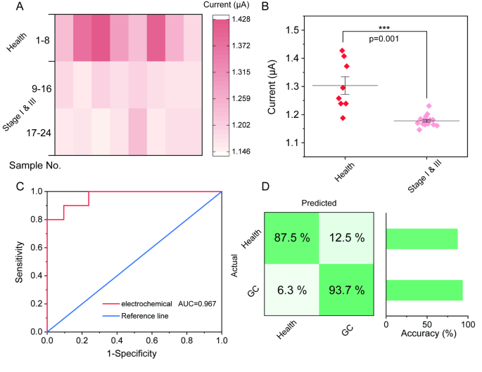
Primarily based on simulated outcomes, scientific samples had been collected to additional consider the sensible utility of the electrochemical biosensor. Human serum samples from 16 GC sufferers at phases I & III and eight damaging management samples had been collected and extracted as scientific samples. The concentrations of miR-19b-3p within the scientific samples had been decided each by classical qRT-PCR and this biosensor (Desk S3). The ends in Fig. 5A and Fig. S8 clearly present that the expression stage of miR-19b-3p in GC stage I & III sufferers is decrease than that in wholesome people (Well being), indicating that miR-19b-3p is downregulated in GC sufferers [15, 16, 34, 35]. In line with the numerous evaluation end result, the proposed electrochemical biosensor can effectively distinguish gastric most cancers (GC) sufferers and wholesome people (Fig. 5B). This conclusion is nice settlement with qRT-PCR outcomes, that are recorded in Fig. S9. Furthermore, the detection efficacy was additionally investigated through the use of a receiver working attribute (ROC) curve (Fig. 5C). Notably, the world beneath the curve (AUC) of this electrochemical biosensor is 0.967. Additional evaluation confirmed that the false optimistic fee was 6.3% and the false damaging fee was 12.5%, proving that the accuracies for classification of Well being versus GC. (Fig. 5D). Above outcomes additional prompt that the proposed electrochemical sensing technique holds an excellent potential utility within the scientific prognosis of gastric most cancers.
Evaluation of miR-19b-3p expression ranges in scientific blood samples by the proposed biosensor. (A) Warmth map of electrochemical sign of this biosensor for miR-19b-3p detection within the blood of Wholesome individuals, GC stage I & III sufferers. (B) Important discount in electrochemical sign for the GC stage I & III sufferers in contrast with wholesome individuals (p = 0.001). (C) ROC evaluation of miR-19b-3p in scientific blood samples by the electrochemical biosensor. (D) Confusion matrix summarizing the GC prognosis ends in a validation cohort

